
















Copyright: ©Author(s) 2026.
World J Diabetes. Jun 15, 2026; 17(6): 118576
Published online Jun 15, 2026. doi: 10.4239/wjd.118576
Published online Jun 15, 2026. doi: 10.4239/wjd.118576
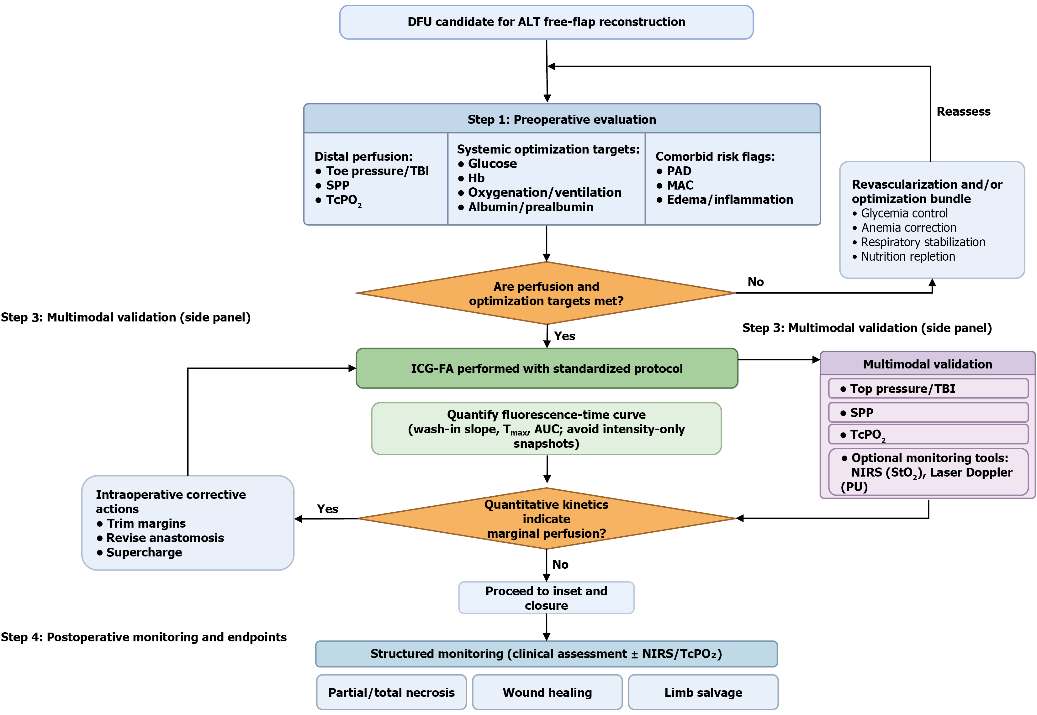
Figure 2 Quantitative, multimodal pathway for anterolateral thigh flap perfusion in diabetic foot ulcer.
This workflow summarizes a stepwise, bundle-based algorithm for diabetic foot ulcer (DFU) candidates undergoing anterolateral thigh (ALT) free-flap reconstruction. Step 1 (preoperative evaluation) integrates distal perfusion testing [toe pressure/toe-brachial index (TBI), skin perfusion pressure (SPP), transcutaneous oxygen tension (TcPO2)], systemic optimization targets [glucose control, hemoglobin (Hb), oxygenation/ventilation, nutritional reserve], and comorbidity risk flags [peripheral artery disease (PAD), medial arterial calcification (MAC), edema/inflammation]. If perfusion and optimization targets are not met, an optimization/revascularization bundle is initiated and reassessed before proceeding. Step 2 (intraoperative perfusion imaging) applies indocyanine green fluorescence angiography (ICG-FA) under a standardized acquisition protocol and prioritizes fluorescence-time curve quantification [e.g., wash-in slope, time-to-peak, area under the curve (AUC)] to avoid intensity-only snapshots. Step 3 (multimodal validation) cross-checks ICG-FA kinetics against distal perfusion metrics and optional monitoring tools (near-infrared spectroscopy [NIRS]/tissue oximetry and laser Doppler perfusion) to adjudicate marginal perfusion. When quantitative kinetics suggest borderline perfusion, targeted intraoperative corrective actions (trim hypoperfused margins, revise anastomosis, supercharging) are implemented; otherwise, the surgeon proceeds to inset/closure. Step 4 (postoperative monitoring and endpoints) emphasizes structured surveillance (clinical assessment with optional near-infrared spectroscopy/TcPO2) and links the pathway to clinically meaningful outcomes, including partial/total necrosis, wound healing, and limb salvage. Tmax: Time to maximum fluorescence intensity; StO2: Tissue oxygen saturation; PU: Perfusion units.
- Citation: Lu PY, Hao ZY, Xing GW, Zhang PF, Liu YS, Li WY, Xu MJ. Improving intraoperative perfusion reliability in anterolateral thigh free flap reconstruction for diabetic foot ulcers. World J Diabetes 2026; 17(6): 118576
- URL: https://www.wjgnet.com/1948-9358/full/v17/i6/118576.htm
- DOI: https://dx.doi.org/10.4239/wjd.118576