
















Copyright: ©Author(s) 2026.
World J Diabetes. Jun 15, 2026; 17(6): 118576
Published online Jun 15, 2026. doi: 10.4239/wjd.118576
Published online Jun 15, 2026. doi: 10.4239/wjd.118576
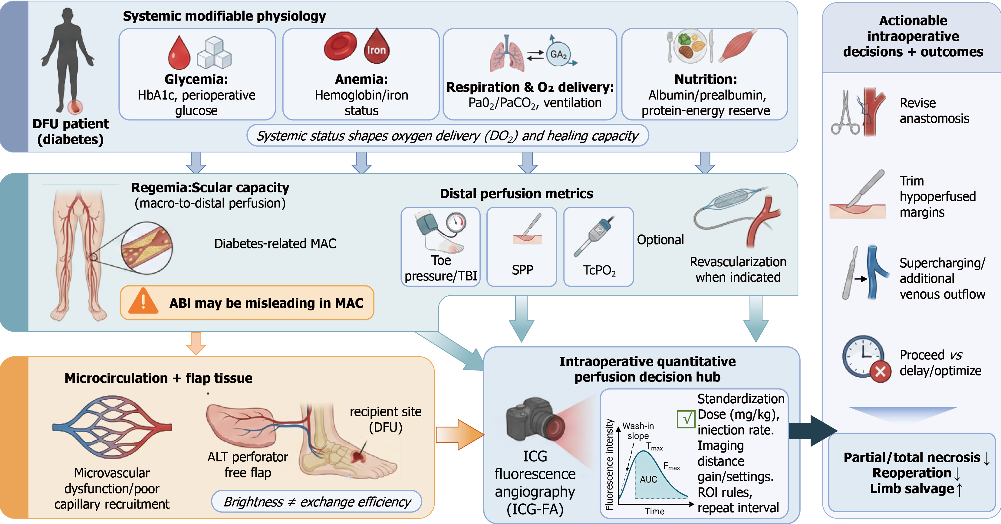
Figure 1 Improving anterolateral thigh free-flap perfusion reliability in diabetic foot ulcers.
This schematic frames anterolateral thigh (ALT) free-flap perfusion in diabetic foot ulcer (DFU) as an integrated continuum linking systemic modifiable physiology - glycemic status [hemoglobin A1c (HbA1c)/perioperative glucose], anemia/iron status, respiration and oxygen delivery (DO2) [arterial partial pressure of carbon dioxide/oxygen (PaCO2/PaO2), ventilation], and nutritional reserve (albumin/prealbumin) - to regional vascular capacity in the setting of diabetes-related medial arterial calcification (MAC), where ankle-brachial index (ABI) may be misleading, and distal perfusion should be characterized using toe pressure/toe-brachial index (TBI), skin perfusion pressure (SPP), and transcutaneous oxygen tension (TcPO2), with revascularization when indicated. These layers converge with microcirculatory dysfunction and impaired capillary recruitment at the flap and recipient-site level (conceptual note: “Brightness ≠ exchange efficiency”). The central intraoperative quantitative perfusion decision hub integrates systemic status, distal perfusion context, and standardized indocyanine green fluorescence angiography (ICG-FA) acquisition, emphasizing fluorescence-time curve kinetics [wash-in slope, time to maximum intensity, maximum fluorescence intensity, area under the curve (AUC)] rather than single intensity snapshots. Outputs are actionable intraoperative strategies (revise anastomosis, trim hypoperfused margins, supercharging/additional venous outflow, proceed vs delay/optimize) to reduce partial/total necrosis and reoperation and improve limb salvage. ROI: Region of interest.
- Citation: Lu PY, Hao ZY, Xing GW, Zhang PF, Liu YS, Li WY, Xu MJ. Improving intraoperative perfusion reliability in anterolateral thigh free flap reconstruction for diabetic foot ulcers. World J Diabetes 2026; 17(6): 118576
- URL: https://www.wjgnet.com/1948-9358/full/v17/i6/118576.htm
- DOI: https://dx.doi.org/10.4239/wjd.118576