
















Copyright: ©Author(s) 2026.
World J Diabetes. May 15, 2026; 17(5): 118275
Published online May 15, 2026. doi: 10.4239/wjd.v17.i5.118275
Published online May 15, 2026. doi: 10.4239/wjd.v17.i5.118275
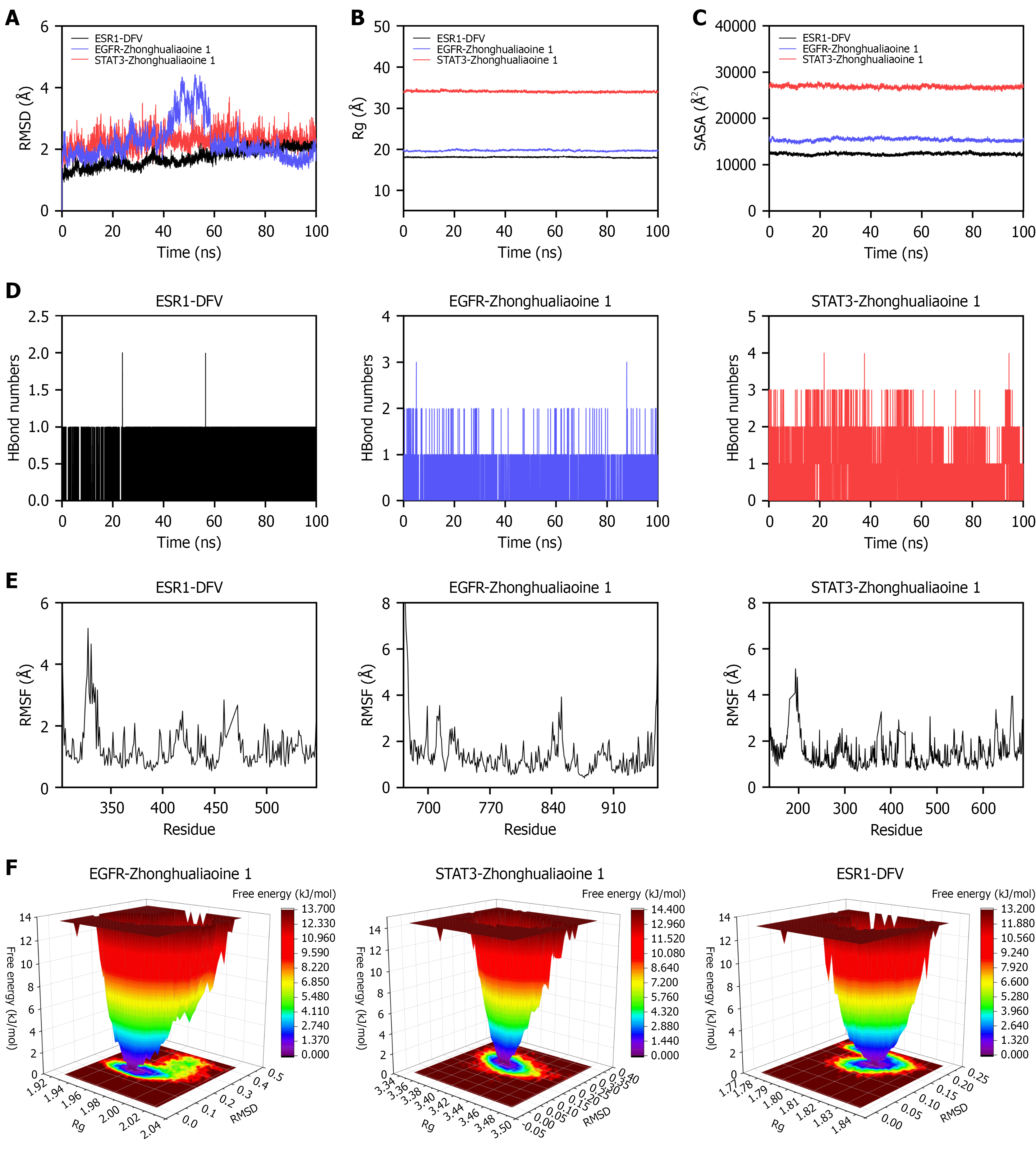
Figure 4 Molecular dynamics simulation of the protein-ligand complexes.
A: Root-mean-square deviation; B: Radius of gyration; C: Solvent-accessible surface area; D: Hydrogen bond count; E: Root-mean-square fluctuation; F: Free energy landscape. Receptors: AMBER14SB force field; ligands: GAFF2 (RESP charges); solvated in TIP3P water box (1 nm edge), 0.15 M NaCl; long-range electrostatics: Particle mesh Ewald method. Molecular dynamics results (GROMACS 2022, 100 nanoseconds, normal pressure and temperature: 310 K, 1 bar) of estrogen receptor 1/diosgenin-3-O-β-D-fructofuranosyl-α-L-rhamnopyranoside, epidermal growth factor receptor/zhonghualiaoine 1, signal transducer and activator of transcription 3/zhonghualiaoine 1 complexes. DFV: Diosgenin-3-O-β-D-fructofuranosyl-α-L-rhamnopyranoside; EGFR: Epidermal growth factor receptor; ESR1: Estrogen receptor 1; Rg: Radius of gyration; RMSD: Root-mean-square deviation; RMSF: Root-mean-square fluctuation; SASA: Solvent-accessible surface area; STAT3: Signal transducer and activator of transcription 3.
- Citation: Li WJ, Xu JJ, Tiang HS, Liu TH, Wu LL. Network pharmacology: Anti-obesity and insulin-sensitizing effects of Danggui-Huangjing herb-pair via epidermal growth factor receptor/phosphatidylinositol 3-kinase/protein kinase B pathway. World J Diabetes 2026; 17(5): 118275
- URL: https://www.wjgnet.com/1948-9358/full/v17/i5/118275.htm
- DOI: https://dx.doi.org/10.4239/wjd.v17.i5.118275