
















Copyright: ©Author(s) 2026.
World J Diabetes. May 15, 2026; 17(5): 117132
Published online May 15, 2026. doi: 10.4239/wjd.v17.i5.117132
Published online May 15, 2026. doi: 10.4239/wjd.v17.i5.117132
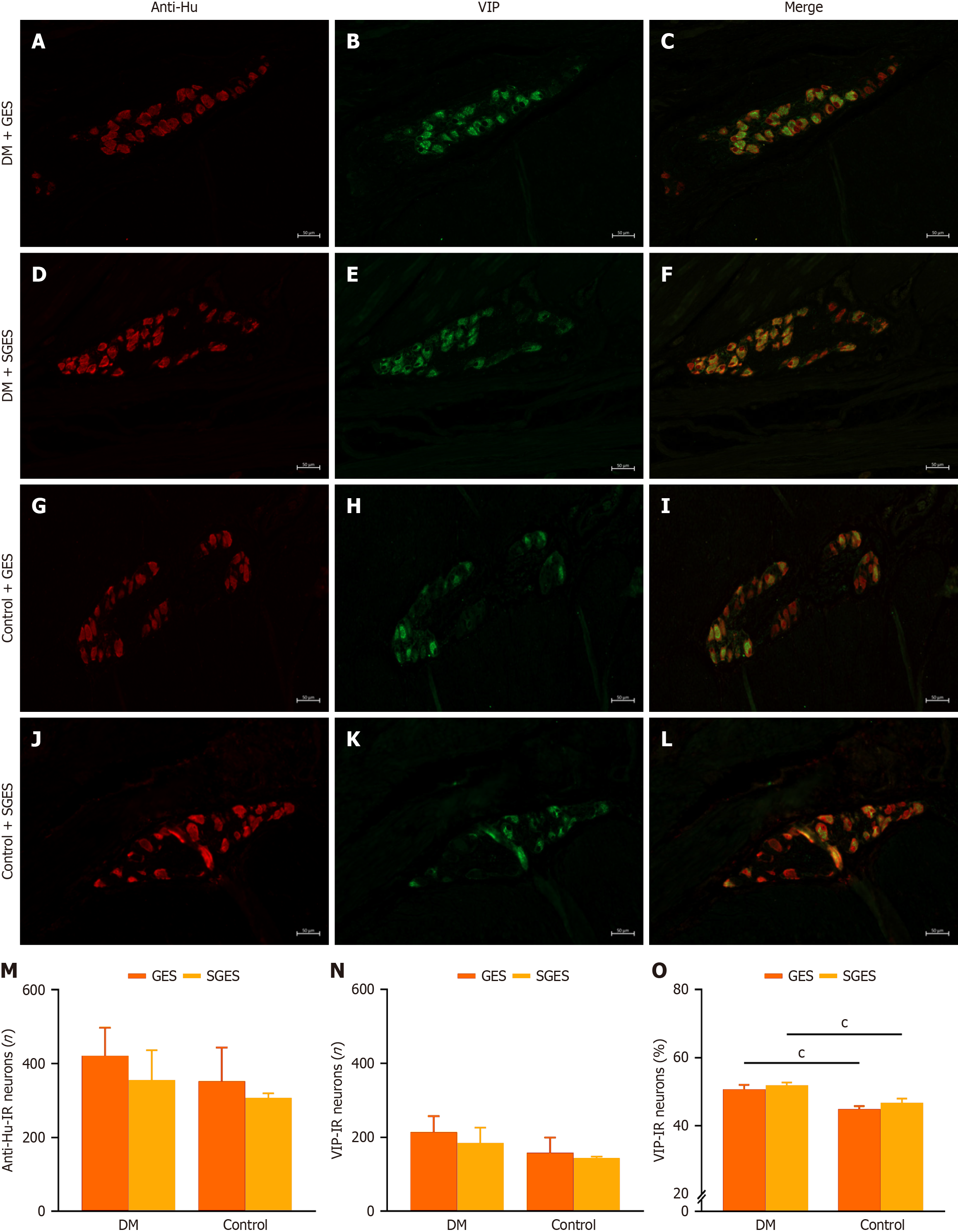
Figure 4 Representative immunofluorescence images of myenteric vasoactive intestinal peptide-immunoreactive positive neurons in full-thickness sections of canine gastric antrum (200 × magnification, scale bar = 50 μm).
A: Representative images of anti-human neuronal protein HuC/HuD-immunoreactive (anti-Hu-IR) labeled neurons in diabetes mellitus (DM) + gastric electrical stimulation (GES); B: Representative images of vasoactive intestinal peptide-immunoreactive (VIP-IR) positive neurons in DM + GES; C: Representative merged images of anti-Hu-IR/VIP-IR in DM + GES; D: Representative images of anti-Hu-IR labeled neurons in DM + sham GES (SGES); E: Representative images of VIP-IR positive neurons in DM + SGES; F: Representative merged images of anti-Hu-IR/VIP-IR in DM + SGES; G: Representative images of anti-Hu-IR labeled neurons in control + GES; H: Representative images of VIP-IR positive neurons in control + GES; I: Representative merged images of anti-Hu-IR/VIP-IR in control + GES; J: Representative images of anti-Hu-IR labeled neurons in control + SGES; K: Representative images of VIP-IR positive neurons in control + SGES; L: Representative merged images of anti-Hu-IR/VIP-IR in control + SGES; M: Number of anti-Hu-IR positive neurons; N: Number of VIP-IR positive neurons; O: Proportion of VIP-IR positive neurons relative to total neurons. The proportion of VIP-IR neurons was significantly higher in the diabetes mellitus canines compared with controls, whether in GES or SGES subgroups (cP < 0.001). Anti-Hu: Anti-human neuronal protein HuC/HuD; DM: Diabetes mellitus; GES: Gastric electrical stimulation; IR: Immunoreactive; SGES: Sham gastric electrical stimulation; VIP: Vasoactive intestinal peptide.
- Citation: Tang P, Wu X, Xi JR, Fu MY, Xue XW, Jiang Y, Zhao Y, Yang HE, Fei GJ, Li XQ, Hu CH, Fang XC. Gastric electrical stimulation ameliorates antral myenteric neurons in streptozotocin-induced diabetic beagle canines. World J Diabetes 2026; 17(5): 117132
- URL: https://www.wjgnet.com/1948-9358/full/v17/i5/117132.htm
- DOI: https://dx.doi.org/10.4239/wjd.v17.i5.117132