
















Copyright: ©Author(s) 2026.
World J Diabetes. Apr 15, 2026; 17(4): 114679
Published online Apr 15, 2026. doi: 10.4239/wjd.v17.i4.114679
Published online Apr 15, 2026. doi: 10.4239/wjd.v17.i4.114679
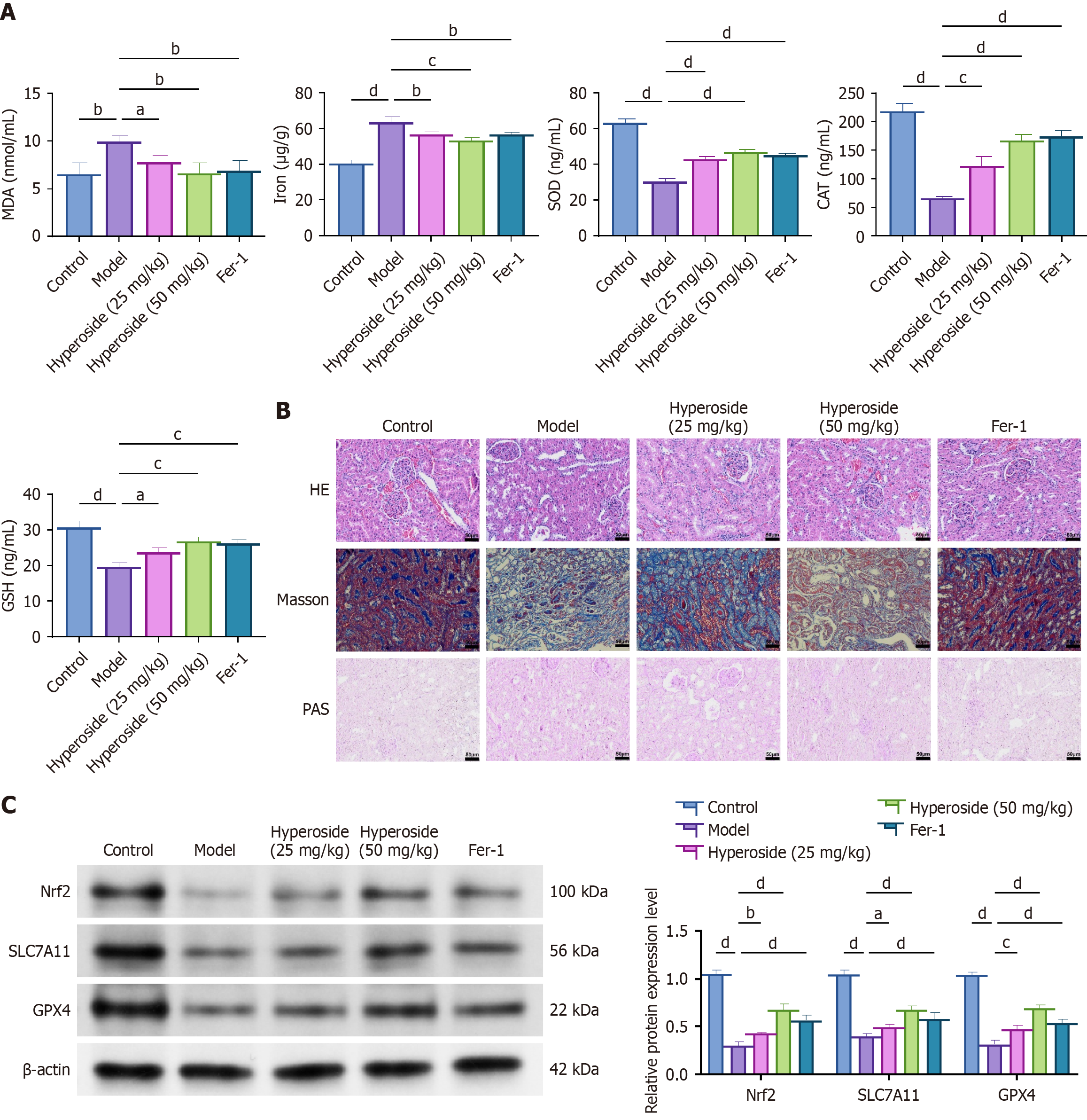
Figure 7 Hyperoside mitigates oxidative stress and glomerular structural damage in diabetic nephropathy rats.
aP < 0.05, bP < 0.01, cP < 0.001, dP < 0.001. One-way analysis of variance + Tukey’s post hoc test; two-sided. Data are expressed as the mean ± SD; n = 6 rats per group. A: Renal malondialdehyde, Fe, superoxide dismutase, catalase, and glutathione; B: Representative hematoxylin and eosin, Masson, and periodic acid-Schiff staining with semi-quantitative scores calculated by two blinded observers; C: Western blot analysis of renal Nrf2, SLC7A11, and GPX4 expression. Fer-1: Ferrostatin-1; MDA: Malondialdehyde; SOD: Superoxide dismutase; CAT: Catalase; GSH: Glutathione; PAS: Periodic acid-Schiff; HE: Hematoxylin and eosin.
- Citation: Liu C, Li Y, Zhang Y, Gao M, Yang SF. Hyperoside attenuates diabetic nephropathy by activating the Nrf2/SLC7A11/GPX4 axis to restrain ferroptosis. World J Diabetes 2026; 17(4): 114679
- URL: https://www.wjgnet.com/1948-9358/full/v17/i4/114679.htm
- DOI: https://dx.doi.org/10.4239/wjd.v17.i4.114679