
















Copyright: ©Author(s) 2026.
World J Diabetes. Apr 15, 2026; 17(4): 114679
Published online Apr 15, 2026. doi: 10.4239/wjd.v17.i4.114679
Published online Apr 15, 2026. doi: 10.4239/wjd.v17.i4.114679
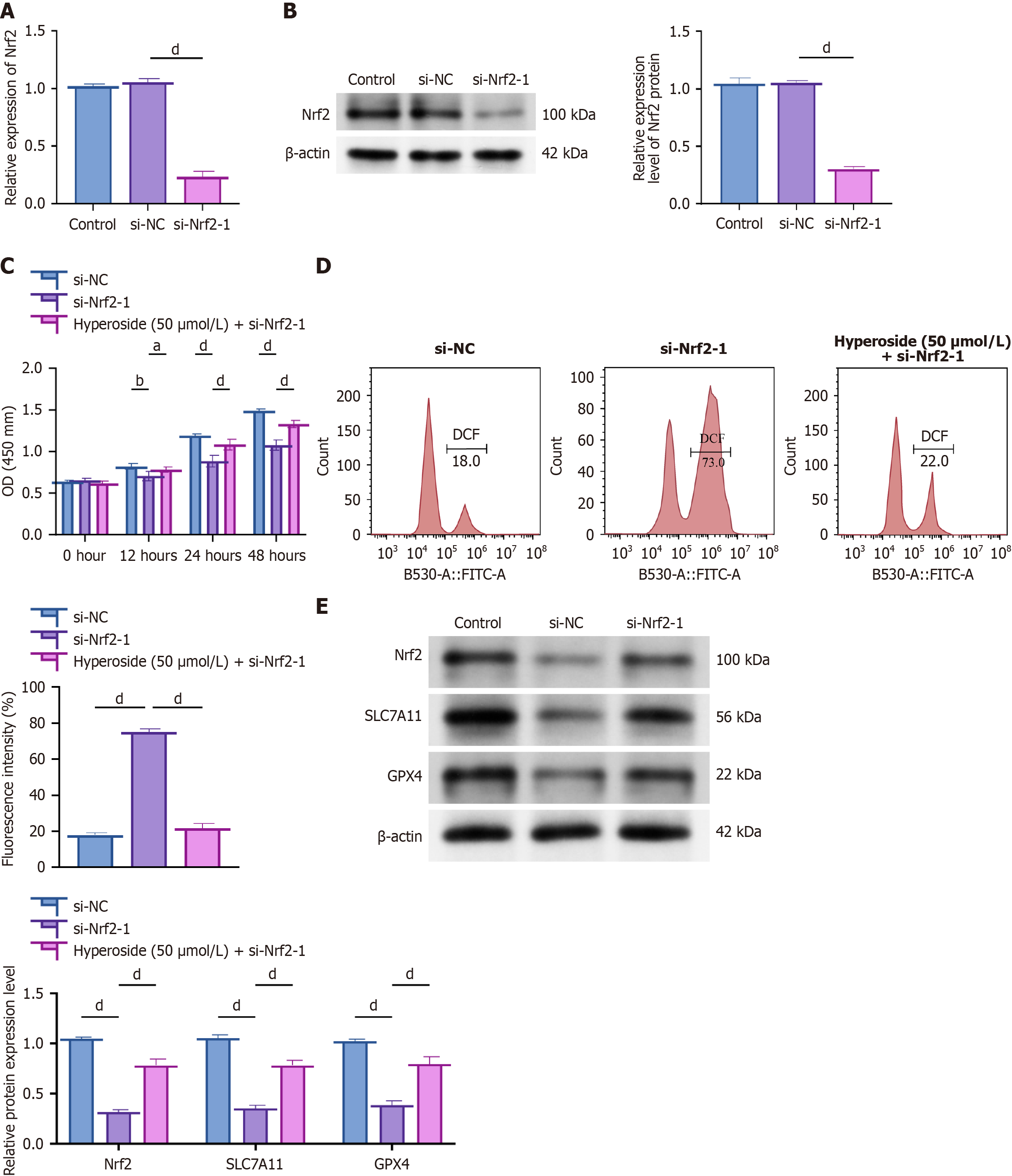
Figure 4 Hyperoside acts via Nrf2 to restrain ferroptosis (siRNA validation).
aP < 0.05, bP < 0.01, dP < 0.001. One-way analysis of variance + Tukey’s post hoc test; two-sided. Data are expressed as the mean ± SD; n = 3 independent experiments. A: Quantitative reverse transcription-polymerase chain reaction confirming Nrf2 knockdown; B: Western blot confirming Nrf2 knockdown; C: Cell Counting Kit-8 time course: Small interfering RNA negative control, small interfering-Nrf2, and small interfering-Nrf2 + hyperoside (50 μM); D: Lipid reactive oxygen species detected using flow cytometry; E: Western blot analysis of Nrf2, SLC7A11, and GPX4 expression. si-NC: Small interfering RNA negative control; OD: Optical density; DCF: 2,7’-dichlorofluorescein; FITC-A: Fluorescein isothiocyanate-area.
- Citation: Liu C, Li Y, Zhang Y, Gao M, Yang SF. Hyperoside attenuates diabetic nephropathy by activating the Nrf2/SLC7A11/GPX4 axis to restrain ferroptosis. World J Diabetes 2026; 17(4): 114679
- URL: https://www.wjgnet.com/1948-9358/full/v17/i4/114679.htm
- DOI: https://dx.doi.org/10.4239/wjd.v17.i4.114679