
















Copyright: ©Author(s) 2026.
World J Diabetes. Apr 15, 2026; 17(4): 114679
Published online Apr 15, 2026. doi: 10.4239/wjd.v17.i4.114679
Published online Apr 15, 2026. doi: 10.4239/wjd.v17.i4.114679
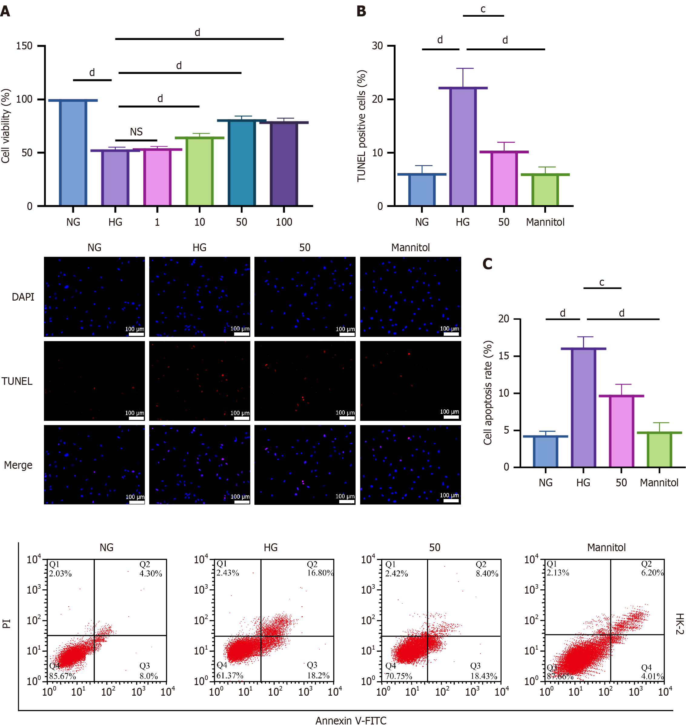
Figure 1 Effects of hyperoside on HK-2 cell viability under high-glucose conditions.
cP < 0.001, dP < 0.001. One-way analyses of variance with Tukey’s post hoc test; two-sided. Data are expressed as the mean ± SD; n = 3 independent experiments. A: Cell Counting Kit-8 assay showing viability with hyperoside (1 μM, 10 μM, 50 μM, and 100 μM) under high glucose (HG) (30 mmol/L); B: Terminal deoxynucleotidyl transferase dUTP nick-end labeling: Normal glucose, HG, and HG + hyperoside (50 μM); quantification of terminal deoxynucleotidyl transferase dUTP nick-end labeling-positive cells; C: Flow cytometry apoptosis rates in the same groups. NG: Normal glucose; HG: High glucose; DAPI: 4’,6-diamidino-2-phenylindole; TUNEL: Terminal deoxynucleotidyl transferase dUTP nick-end labeling; FITC: Fluorescein isothiocyanate.
- Citation: Liu C, Li Y, Zhang Y, Gao M, Yang SF. Hyperoside attenuates diabetic nephropathy by activating the Nrf2/SLC7A11/GPX4 axis to restrain ferroptosis. World J Diabetes 2026; 17(4): 114679
- URL: https://www.wjgnet.com/1948-9358/full/v17/i4/114679.htm
- DOI: https://dx.doi.org/10.4239/wjd.v17.i4.114679