
















Copyright: ©Author(s) 2026.
World J Diabetes. Mar 15, 2026; 17(3): 115433
Published online Mar 15, 2026. doi: 10.4239/wjd.v17.i3.115433
Published online Mar 15, 2026. doi: 10.4239/wjd.v17.i3.115433
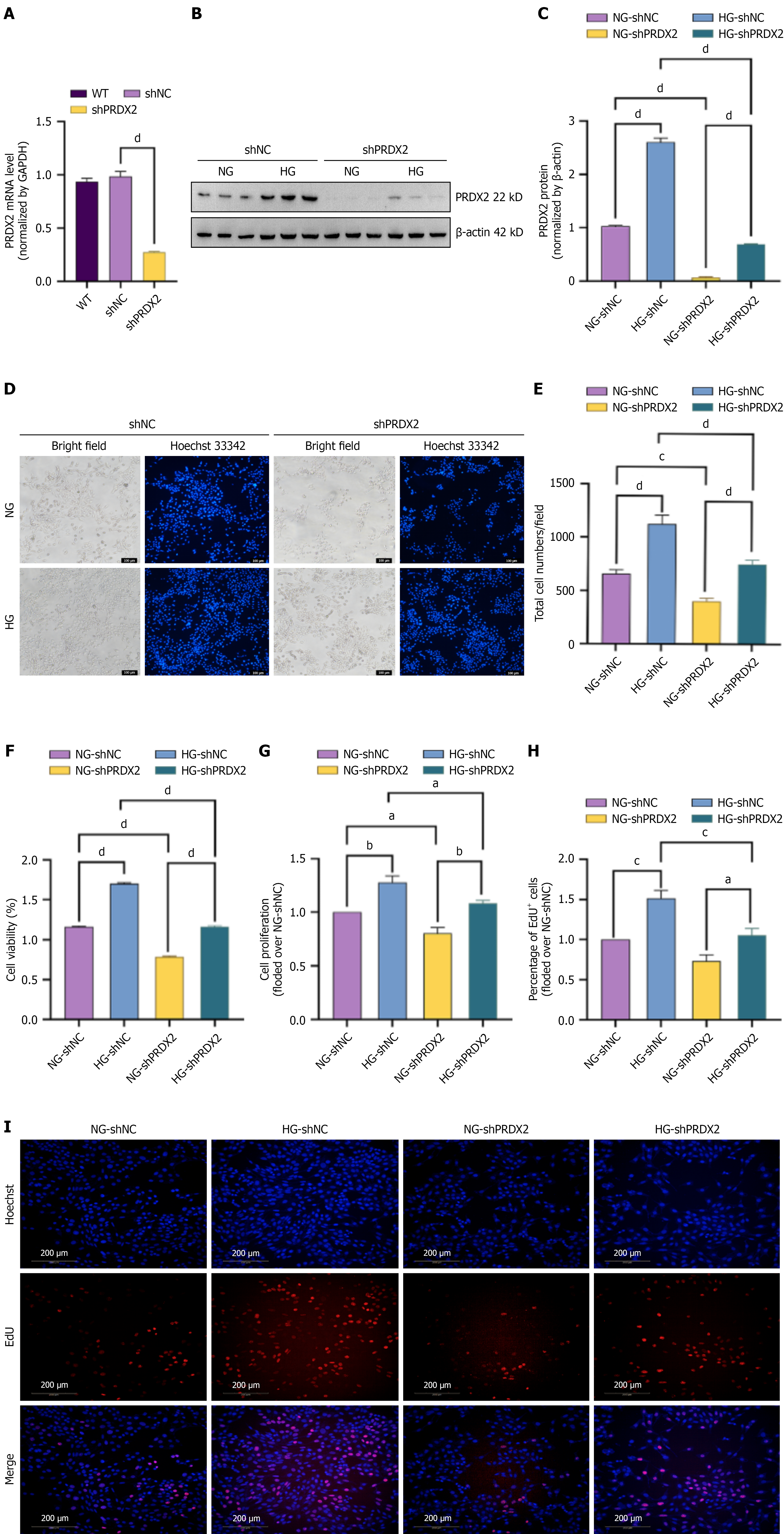
Figure 4 PPDX2 silencing attenuates high glucose-induced Müller cell proliferation.
rMC-1 cells transfected with control (shNC) or PRDX2 shRNA (shPRDX2) before being subjected to high glucose or normal glucose for 48 hours. A: mRNA expression of PRDX2 measured by quantitative polymerase chain reaction in wild-type, shNC, and shPRDX2 rMC-1 cells; B and C: Western blotting analysis of PRDX2 in rMC-1 cells. The bar histogram is representative of measurements of the gray values of PRDX2/β-actin (C); D and E: Hoechst staining was used to measure the total cell number (magnification: 100 ×; scale bars: 100 μm; blue: Hoechst); F: Cell viability as measured by CCK8 assay; G: Cell proliferation was measured by the CyQUANT NF cell proliferation assay kit; H and I: Cell proliferation detected by EdU staining (magnification: 200 ×; scale bars: 200 μm; red: EdU; blue: Hoechst). Bar histogram represents the quantitative results of rMC-1 cell proliferation rate under EdU staining (H). All values are presented as means ± SEM. Each experiment was independently repeated 3-5 times. P value for trends assessed using ordinary one-way analysis of variance. aP < 0.05; bP < 0.01; cP < 0.001; dP < 0.0001. WT: Wild type; NG: Normal glucose; HG: High glucose.
- Citation: Du XL, Ouyang S, Wang YS, Mao YS, Ren BC, Yu WH. PRDX2 silencing alleviates reactive hyperplasia of Müller glial cells in diabetic retinopathy by inhibiting the RhoA/ROCK signaling pathway. World J Diabetes 2026; 17(3): 115433
- URL: https://www.wjgnet.com/1948-9358/full/v17/i3/115433.htm
- DOI: https://dx.doi.org/10.4239/wjd.v17.i3.115433