
















Copyright: ©Author(s) 2026.
World J Diabetes. Mar 15, 2026; 17(3): 113249
Published online Mar 15, 2026. doi: 10.4239/wjd.v17.i3.113249
Published online Mar 15, 2026. doi: 10.4239/wjd.v17.i3.113249
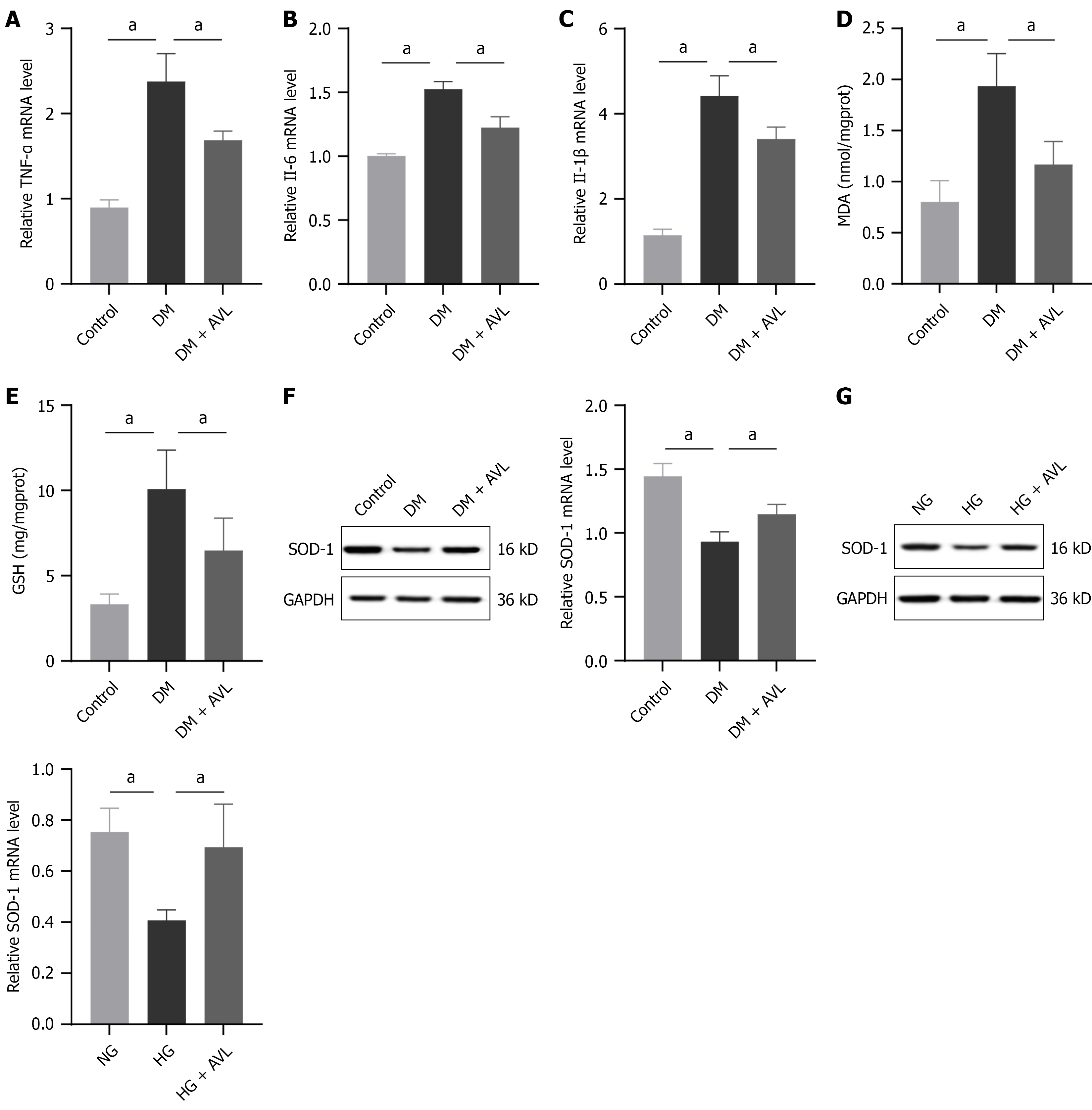
Figure 3 Amomum villosum Lour.
treatment reduced inflammation and oxidative stress in the dorsal root ganglion neurons of diabetic rats. The type 1 diabetes mellitus (DM) rat model was established in week 1. Starting from week 2, Amomum villosum Lour. (AVL; 600 mg/kg/day) was administered intraperitoneally once daily until the end of the experiment. The total study duration was 5 weeks. A-C: MRNA expression levels of tumor necrosis factor alpha (TNF-α), interleukin 6 (IL-6), and IL-1β in dorsal root ganglion (DRG) tissue were analyzed by quantitative PCR; D and E: Levels of malondialdehyde (MDA) and glutathione (GSH) in DRG tissue were detected by the enzyme-linked immunosorbent assay; F: Protein levels of superoxide dismutase 1 (SOD-1) in DRG tissue were analyzed by Western blotting; G: For 24 hours, normal glucose (NG; 5.5 mmol/L) and high glucose (HG; 50 mmol/L) were administered as pretreatments to primary DRG neurons, and AVL treatment was added in the last 12 hours. The protein levels of SOD-1 in primary DRG neurons were detected by Western blotting. aP < 0.05.
- Citation: Kou DQ, Liu MJ, Gao XL, Guo FR. Amomum villosum extract alleviates diabetic neuropathy via phosphoinositide 3-kinase/AKT-mediated antioxidative and anti-apoptotic effects. World J Diabetes 2026; 17(3): 113249
- URL: https://www.wjgnet.com/1948-9358/full/v17/i3/113249.htm
- DOI: https://dx.doi.org/10.4239/wjd.v17.i3.113249