
















Copyright: ©Author(s) 2026.
World J Diabetes. Mar 15, 2026; 17(3): 112177
Published online Mar 15, 2026. doi: 10.4239/wjd.v17.i3.112177
Published online Mar 15, 2026. doi: 10.4239/wjd.v17.i3.112177
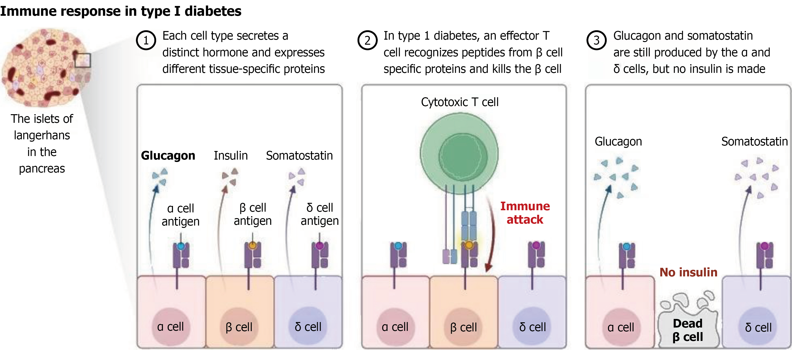
Figure 2 Autoimmune destruction of pancreatic β cells in type 1 diabetes.
The diagram illustrates the pathophysiology of type 1 diabetes mellitus, showing the progressive autoimmune attack on insulin-producing β cells within the pancreatic islets of Langerhans (left panel). The normal pancreatic islet architecture contains α cells (producing glucagon), β cells (producing insulin), and δ cells (producing somatostatin) (middle panel). In type 1 diabetes, autoreactive cytotoxic T cells recognize β cell-specific antigens and mount an immune attack against insulin-producing cells, whereas α and δ cells remain largely unaffected (right panel). The end result of this autoimmune process is the selective destruction of β cells, leading to complete insulin deficiency, whereas glucagon and somatostatin production from α and δ cells continues. This autoimmune-mediated β-cell destruction results in the characteristic hyperglycemia and insulin dependence observed in type 1 diabetes patients. This figure was created by BioRender.com (Supplementary material).
- Citation: Chen X, Zhang LL, Zhou Y, Dong DX, Qian XY. Mitochondrial autophagy in diabetes-related cognitive decline and skin ulcers: Mechanistic insights and therapeutic implications. World J Diabetes 2026; 17(3): 112177
- URL: https://www.wjgnet.com/1948-9358/full/v17/i3/112177.htm
- DOI: https://dx.doi.org/10.4239/wjd.v17.i3.112177