
















©The Author(s) 2026.
World J Diabetes. Feb 15, 2026; 17(2): 113992
Published online Feb 15, 2026. doi: 10.4239/wjd.v17.i2.113992
Published online Feb 15, 2026. doi: 10.4239/wjd.v17.i2.113992
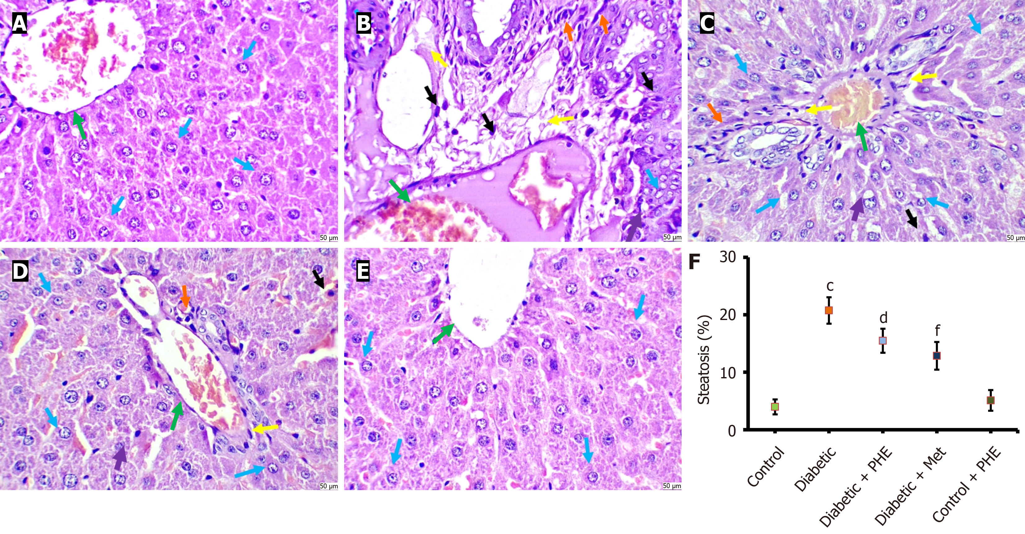
Figure 7 Histopathological changes in the rat liver tissue after 8 weeks of polyherbal extract treatment, with semi-quantitative data as steatosis percentage.
A: Control. No evidence of cellular disintegration, pyknosis, apoptosis, central vein congestion or nuclear morphology was found (green arrow). Furthermore, the sections exhibited normal and trabecular-arranged hepatocytes, which were polygonal in shape (indicated by the blue arrow). The cytoplasm was clear, and the nuclei were normal/round/oval; no fatty changes, inflammatory infiltrates or congestion in the central vein were observed; B: Diabetic. The section showed significantly damaged hepatocytes (indicated by the blue arrow). High cellular density, dense cytoplasm and significant fatty changes (yellow arrow) and vacuolation (purple arrow) were seen. Pyknosis (black arrow) and central vein congestion (green arrow), along with inflammatory infiltrates (orange arrow), were seen. The micromorphological characteristics were suggestive of severe hepatic toxicity; C: Diabetic + polyherbal extract. The section showed mild to moderate histological damage to the liver tissue. Moderate congestion was seen (green arrow). The section showed mildly damaged hepatocytes (blue arrow), mild vacuolation (purple arrow), mild pyknosis (black arrow), moderate fatty changes (yellow arrow) and moderate inflammatory infiltrates (orange arrow); D: Diabetic + metformin. The section showed mild histological damage to the liver tissue. Minimal congestion was seen (green arrow). The section showed normal hepatocytes (indicated by the blue arrow). Minimal fatty changes (yellow arrow), minimal vacuolation (purple arrow) and minimal pyknosis (black arrow) along with minimal inflammatory infiltrates (orange arrow) were seen; E: Control + polyherbal extract. The section showed the normal histological appearance of the liver tissue. No evidence of cellular disintegration, pyknosis, apoptosis, central vein congestion or nuclear morphology was found (green arrow). The section also showed normal and trabecular-arranged hepatocytes, which were polygonal in shape (indicated by the blue arrow). The cytoplasm was clear, and the nuclei were normal/round/oval; no fatty changes, inflammatory infiltrates or congestion in the central vein were seen (haematoxylin and eosin, 400 ×; scale bar 50 μm); F: Semi-quantitative analysis of hepatic steatosis percentage in different experimental groups. cP < 0.001 vs control group, dP < 0.05 vs diabetic group, fP < 0.001 vs diabetic group. Met: Metformin; PHE: Polyherbal extract.
- Citation: Kausar MA, Parveen K, Anwar S, Khan YS, Saleh AA, Ahmed MAA, Siddiqui WA, Parvez S. Neuroprotective potential of a plant-based intervention in diabetic neuropathy: Biochemical and behavioral insights from a streptozotocin-induced rat model. World J Diabetes 2026; 17(2): 113992
- URL: https://www.wjgnet.com/1948-9358/full/v17/i2/113992.htm
- DOI: https://dx.doi.org/10.4239/wjd.v17.i2.113992