
















Copyright: ©Author(s) 2026.
World J Diabetes. Nov 15, 2026; 17(11): 125802
Published online Nov 15, 2026. doi: 10.4239/wjd.125802
Published online Nov 15, 2026. doi: 10.4239/wjd.125802
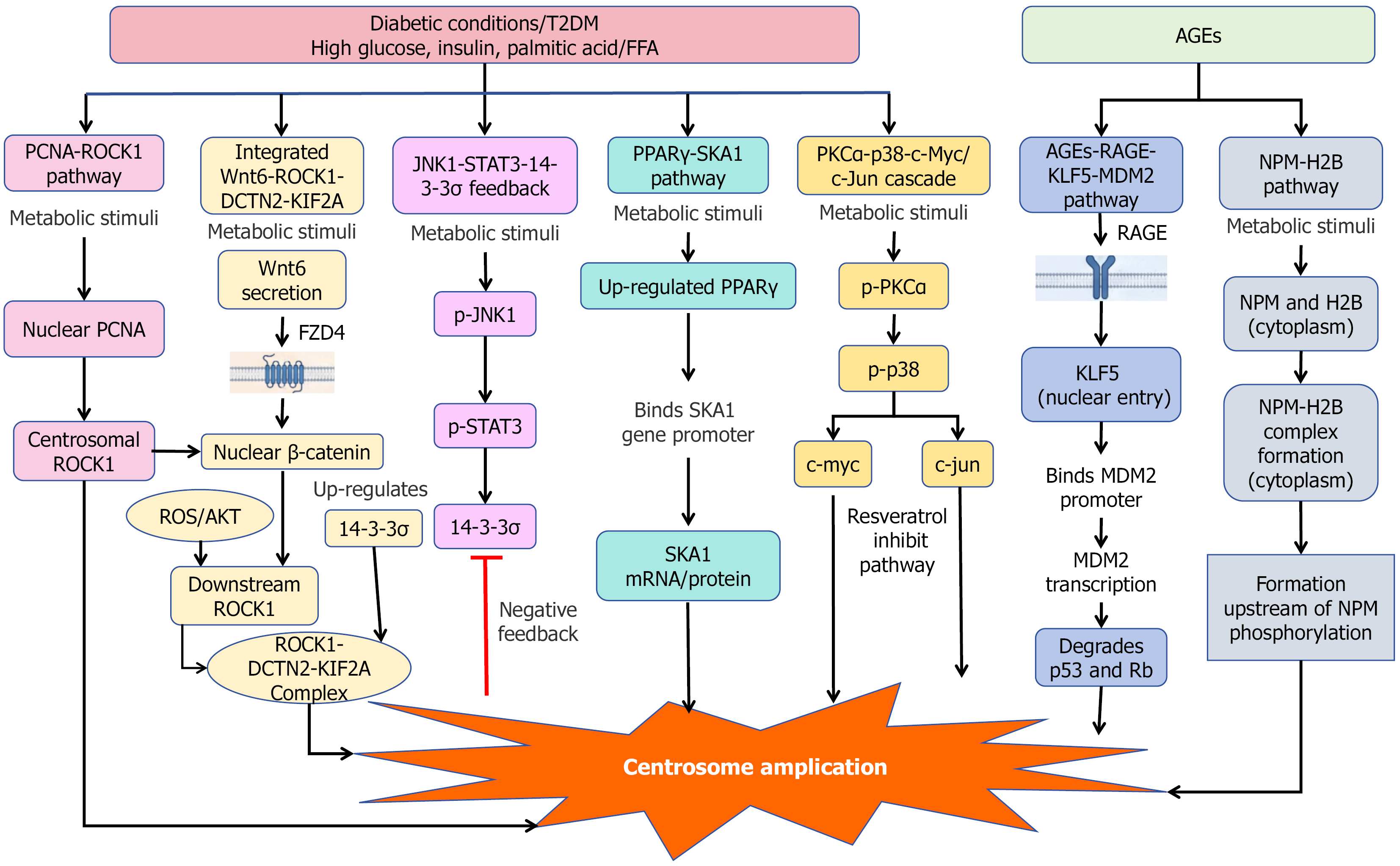
Figure 1 Integrated signaling networks mediating centrosome amplification under diabetic conditions.
Under the pathological microenvironment of type 2 diabetes mellitus, multiple metabolic stressors - including high glucose, insulin, palmitic acid/free fatty acids, and advanced glycation end products - activate converging signaling cascades that collectively drive centrosome amplification (CA). The core pro-CA axes include: (1) The proliferating cell nuclear antigen-Rho-associated protein kinase 1 (ROCK1) axis, which promotes assembly of the centrosomal ROCK1/14-3-3σ complex via upstream reactive oxygen species/protein kinase B signaling; (2) The Wnt6/plasma membrane receptor frizzled 4/β-catenin pathway, in which secreted Wnt6 binds to the membrane receptor plasma membrane receptor frizzled 4 and activates nuclear β-catenin to transcriptionally upregulate ROCK1 and 14-3-3σ; (3) Diabetic stimuli promote centrosome amplification by enhancing ROCK1 binding to dynactin subunit 2 and kinesin family member 2A; (4) The protein kinase C alpha-p38 mitogen-activated protein kinase-cellular myelocytomatosis oncogene/cellular Jun proto-oncogene transcriptional cascade, which activates the transcription of centrosome duplication-related genes; (5) The nucleophosmin-histone H2B complex pathway, where the nuclear nucleophosmin-histone H2B complex translocates to the centrosome and disrupts centrosome duplication control; (6) The peroxisome proliferator-activated receptor gamma-spindle-and kinetochore-associated complex subunit 1 regulatory axis, which modulates centrosome homeostasis via transcriptional upregulation of spindle-and kinetochore-associated complex subunit 1; (7) the advanced glycation end products-receptor for advanced glycation end-products-Krüppel-like factor 5-mouse double minute 2 signaling axis, which facilitates CA through mouse double minute 2-mediated degradation of p53 and Rb; and (8) The c-Jun N-terminal kinase 1-signal transducer and activator of transcription 3-14-3-3σ pathway serves as an endogenous negative feedback loop to restrain excessive CA. Symbols: Solid arrow (→) = promotion, activation, or transcriptional upregulation; blunt-ended line (⊣) = inhibition. T2DM: Type 2 diabetes mellitus; FFA: Free fatty acids; PCNA: Proliferating cell nuclear antigen; ROCK1: Rho-associated coiled-coil-containing protein kinase 1; DCTN2: Dynactin subunit 2; KIF2A: Kinesin family member 2A; JNK1: C-Jun N-terminal kinase 1; STAT3: Signal transducer and activator of transcription 3; PPARγ: Peroxisome proliferator-activated receptor gamma; SKA1: Spindle-and kinetochore-associated complex subunit 1; PKCα: Protein kinase C alpha; c-Myc: Cellular myelocytomatosis oncogene; c-Jun: Cellular Jun proto-oncogene; AGEs: Advanced glycation end-products; RAGE: Receptor for advanced glycation end-products; KLF5: Krüppel-like factor 5; MDM2: Mouse double minute 2; NPM-H2B: Nucleophosmin-histone H2B complex; p-JNK1: Phosphorylated c-Jun N-terminal kinase 1; p-STAT3: Phosphorylated signal transducer and activator of transcription 3; p-PKCα: Phosphorylated protein kinase C alpha; Rb: Retinoblastoma protein; NPM: Nucleophosmin; AGEs: Advanced glycation end-products.
- Citation: Zhang YH, Song ZZ, Zhang S. Centrosome amplification in type 2 diabetes mellitus: Putative mechanisms, clinical research implications, and investigational therapeutic perspectives. World J Diabetes 2026; 17(11): 125802
- URL: https://www.wjgnet.com/1948-9358/full/v17/i11/125802.htm
- DOI: https://dx.doi.org/10.4239/wjd.125802