
















Copyright: ©Author(s) 2026.
World J Diabetes. Nov 15, 2026; 17(11): 124042
Published online Nov 15, 2026. doi: 10.4239/wjd.124042
Published online Nov 15, 2026. doi: 10.4239/wjd.124042
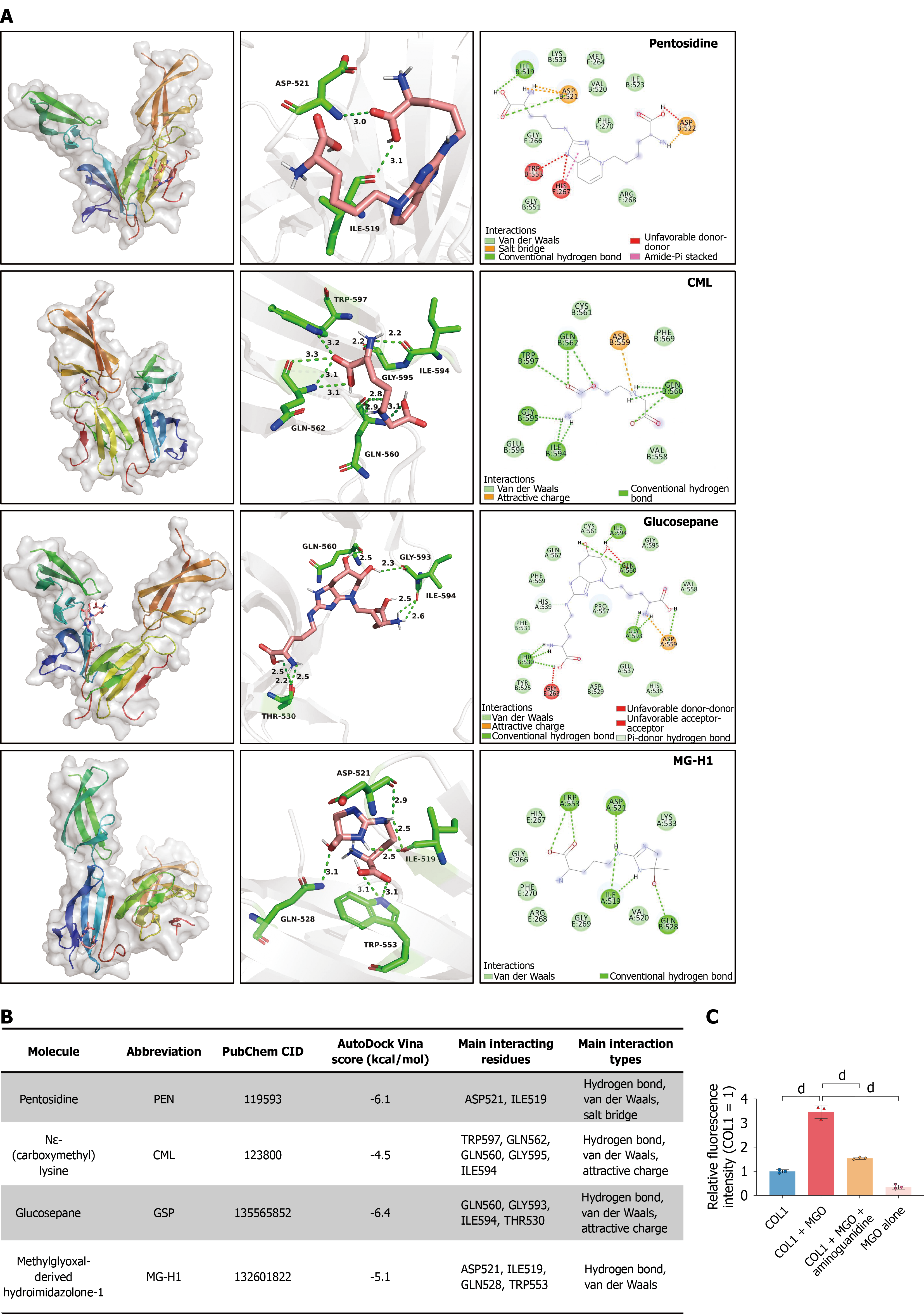
Figure 2 Exploratory non-covalent docking within a fibronectin-collagen peptide complex and methylglyoxal-induced collagen fluorescence.
A: Top-ranked docking poses of pentosidine, Nε-(carboxymethyl)lysine (CML), glucosepane, and methylglyoxal-derived hydroimidazolone-1 (MG-H1) within the crystal structure of a low-affinity collagen site in complex with fibronectin type I domains 8 and 9 (Protein Data Bank accession code: 3GXE). Each row shows the overall predicted pose, local three-dimensional contacts, and two-dimensional interaction diagram. The labeled contacts were predominantly located in fibronectin chains A and B. Specifically, pentosidine contacted aspartate (Asp) 521 and isoleucine (Ile) 519 in chain B; CML contacted tryptophan (Trp) 597, glutamine (Gln) 562, Gln560, glycine (Gly) 595, and Ile594 in chain B; glucosepane contacted Gln560, Gly593, and Ile594 in chain A and threonine 530 in chain B; and MG-H1 contacted Asp521, Ile519, and Trp553 in chain A and Gln528 in chain B. The receptor contained short collagen α1(I)-derived peptides but did not represent full-length fibrillar collagen I (COL1); B: Summary of the AutoDock Vina scores, PubChem identifiers, predicted contacts, and interaction types. More negative Vina scores were used only to rank poses within this model and were not interpreted as experimentally measured binding affinities. Each ligand underwent one docking run, and the nine generated poses were alternative solutions rather than independent replicates. Standard docking did not model glycation chemistry or covalent crosslink formation; C: Advanced glycation end product-related fluorescence in a cell-free COL1-methylglyoxal (MGO) system. Rat-tail COL1 gels (1 mg/mL; 50 μL/well) were overlaid with 100 μL phosphate-buffered saline containing 10 mmol/L MGO, with or without 20 mmol/L aminoguanidine, and incubated at 37 °C for 5 days. MGO increased COL1-associated fluorescence, whereas aminoguanidine reduced this increase. Fluorescence was measured at excitation and emission wavelengths of 370 nm and 440 nm and normalized to the COL1-only group. The MGO-only group controlled for background fluorescence. Data are presented as the mean ± SD from three reaction sets, each measured in three technical wells. Technical-well values were averaged within each reaction set. Multiple-group comparisons were performed using one-way analysis of variance followed by Tukey’s multiple-comparisons test. Ganagliflozin proline was not included in this experiment. The fluorescence assay did not identify specific glycation adducts, modified COL1 residues, or covalent crosslinks. dP < 0.0001. CML: Nε-(carboxymethyl)lysine; MG-H1: Methylglyoxal-derived hydroimidazolone-1; MGO: Methylglyoxal; COL1: Collagen I.
- Citation: Li ZP, Li H, Xu JL, Liu YJ, Li D, Wang P, Xiang L, Xu SY, Duan CZ, Wang SX, Sun JK, Zhang CJ, He DJ, Luo C. Ganagliflozin proline attenuates osteogenic dysfunction and inflammatory signaling during exposure to advanced glycation end products in murine osteoblasts. World J Diabetes 2026; 17(11): 124042
- URL: https://www.wjgnet.com/1948-9358/full/v17/i11/124042.htm
- DOI: https://dx.doi.org/10.4239/wjd.124042