
















Copyright: ©Author(s) 2026.
World J Diabetes. Nov 15, 2026; 17(11): 121630
Published online Nov 15, 2026. doi: 10.4239/wjd.121630
Published online Nov 15, 2026. doi: 10.4239/wjd.121630
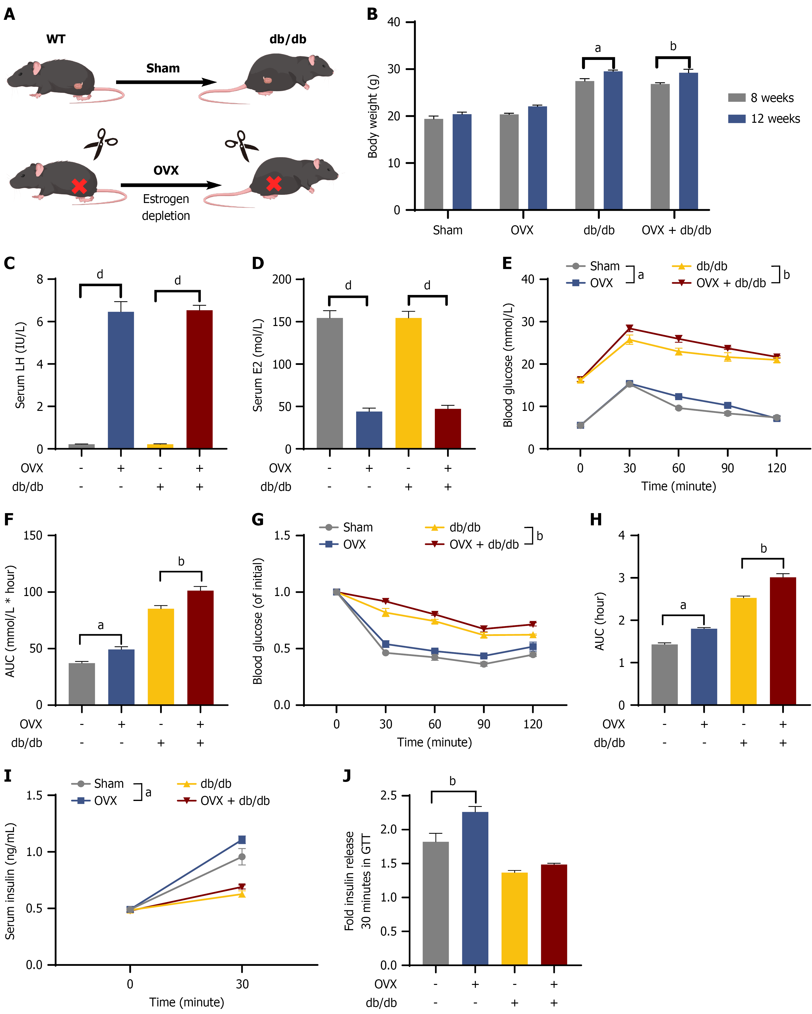
Figure 7 High-endogenous-gonadotropin-releasing hormone milieu is associated with β-cell compensation in vivo.
A: Experimental workflow illustrating the grouping and surgical interventions (sham and ovariectomy) in wild-type and db/db mice; B: Progressive body weight trajectories across the four experimental cohorts; C and D: Serum concentrations of luteinizing hormone (C) and estradiol (D), confirming the successful establishment of the high-gonadotropin-releasing hormone and hypoestrogenic model; E-H: Systemic glucose and insulin homeostasis assessment: Representative curves and corresponding areas under the curve for intraperitoneal glucose tolerance tests (E and F) and insulin tolerance tests (G and H); I and J: Functional evaluation of β-cell compensation: Serum insulin concentrations during the intraperitoneal glucose tolerance tests (I); fold change in insulin secretion (30 minutes vs 0 minute post-glucose challenge), reflecting augmented secretory responsiveness in ovariectomy mice relative to sham controls (J). Data are expressed as the mean ± SEM (n = 8-10 animals per group). aP < 0.05, bP < 0.01, and dP < 0.0001. OVX: Ovariectomy; WT: Wild-type; AUC: Area under the curve.
- Citation: Dai XJ, Gao SC, Chen PT, Yang L, Kong CC, Ye CY, Xu ZX, Hu YX, He CY, Zuo ZH, Wang W. Postmenopausal high-gonadotropin-releasing hormone environment mediates pancreatic β-cell compensation via the cAMP-PKA-PDX1 pathway. World J Diabetes 2026; 17(11): 121630
- URL: https://www.wjgnet.com/1948-9358/full/v17/i11/121630.htm
- DOI: https://dx.doi.org/10.4239/wjd.121630