
















Copyright: ©Author(s) 2026.
World J Diabetes. Nov 15, 2026; 17(11): 121630
Published online Nov 15, 2026. doi: 10.4239/wjd.121630
Published online Nov 15, 2026. doi: 10.4239/wjd.121630
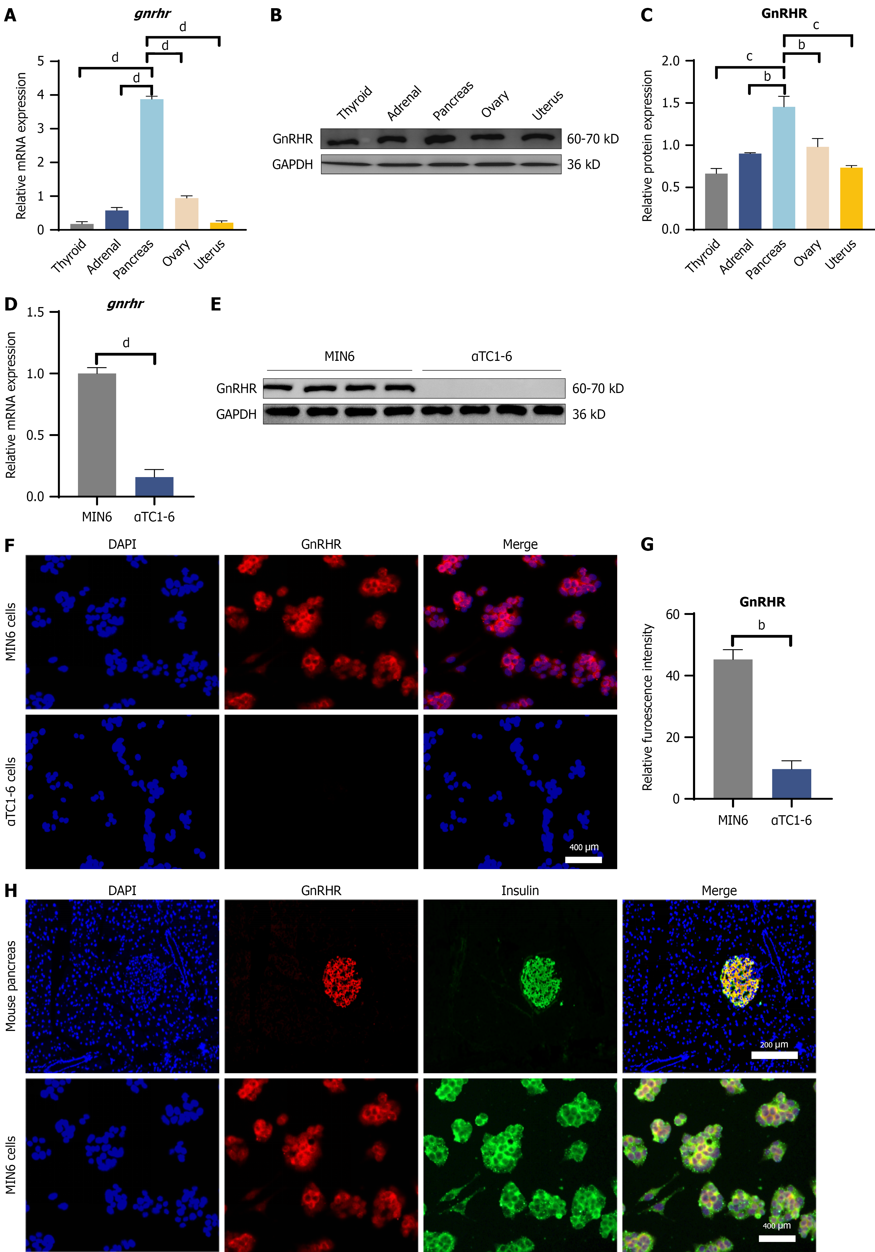
Figure 1 Tissue distribution and β-cell-specific localization of the gonadotropin-releasing hormone receptor.
A-C: Representative immunoblots and quantitative analysis of gonadotropin-releasing hormone receptor (GnRHR) protein expression in the murine pancreas compared with reproductive tissues (ovary and uterus). GAPDH was utilized as the loading control. bP < 0.01 vs ovary or adrenal, cP < 0.001 vs thyroid or uterus, and dP < 0.0001 vs thyroid, adrenal, ovary or uterus; D-G: Comparative analysis of GnRHR expression in murine β-cell (MIN6) vs α-cell (αTC1-6) lines. bP < 0.01 vs αTC1-6, dP < 0.0001 vs αTC1-6. H: Representative dual-immunofluorescence images displaying the colocalization of GnRHR (red) and insulin (green) within pancreatic islets. Nuclei were visualized through DAPI counterstaining (blue). Scale bar = 200 μm. Statistical analysis: Data are expressed as the mean ± SEM from three independent experiments. GnRHR: Gonadotropin-releasing hormone receptor.
- Citation: Dai XJ, Gao SC, Chen PT, Yang L, Kong CC, Ye CY, Xu ZX, Hu YX, He CY, Zuo ZH, Wang W. Postmenopausal high-gonadotropin-releasing hormone environment mediates pancreatic β-cell compensation via the cAMP-PKA-PDX1 pathway. World J Diabetes 2026; 17(11): 121630
- URL: https://www.wjgnet.com/1948-9358/full/v17/i11/121630.htm
- DOI: https://dx.doi.org/10.4239/wjd.121630