
















Copyright: ©Author(s) 2026.
World J Diabetes. Oct 15, 2026; 17(10): 124847
Published online Oct 15, 2026. doi: 10.4239/wjd.124847
Published online Oct 15, 2026. doi: 10.4239/wjd.124847
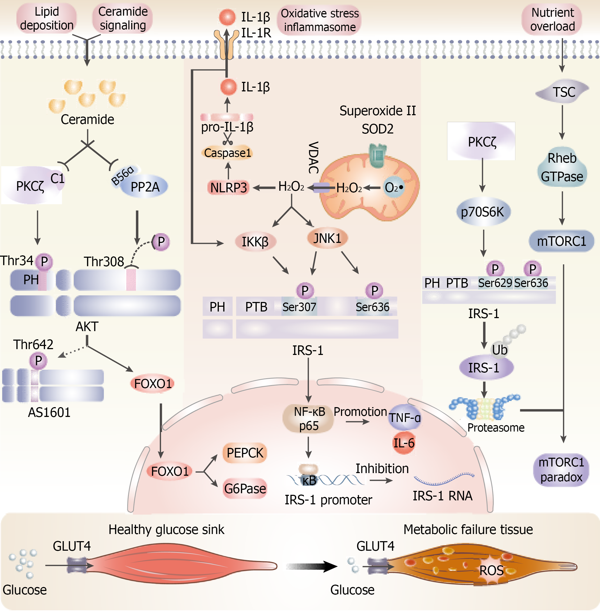
Figure 4 Mechanisms underlying ceramide-induced and reactive oxygen species-induced insulin resistance in skeletal muscle involve multiple interconnected pathways.
Ceramide impairs protein kinase B activity through inhibitory phosphorylation mediated by PKCζ and dephosphorylation facilitated by PP2A, thereby disrupting GLUT4 translocation. Concurrently, mitochondrial hydrogen peroxide (H2O2) activates IKKβ and JNK1, which promote inhibitory phosphorylation and subsequent degradation of insulin receptor substrate-1 (IRS-1). nuclear factor kappa-B suppresses IRS-1 gene transcription. Reactive oxygen species further stimulate the NLRP3 inflammasome, leading to interleukin-1β production that perpetuates inflammatory signaling. Nutrient excess activates the mechanistic target of rapamycin complex 1. PKCζ-mediated activation of p70S6K induces feedback phosphorylation of IRS-1. These coordinated molecular events convert skeletal muscle from an efficient glucose-utilizing tissue into one characterized by chronic insulin resistance and metabolic dysfunction. TNF-α: Tumor necrosis factor-alpha; IL: Interleukin; AKT: Protein kinase B; FOXO: Forkhead box O transcription factors; SOD: Superoxide dismutase; IRS-1: Insulin receptor substrate-1; TSC: Tuberous sclerosis complex; mTORC1: Mechanistic target of rapamycin complex 1.
- Citation: Xie XL, Cao HY, Wang ZT, Chen HR, Zhou LS, Qian T, Han ZY. Pathophysiological mechanisms and risk factors linking type 2 diabetes mellitus and sarcopenia: A narrative review. World J Diabetes 2026; 17(10): 124847
- URL: https://www.wjgnet.com/1948-9358/full/v17/i10/124847.htm
- DOI: https://dx.doi.org/10.4239/wjd.124847