
















Copyright: ©Author(s) 2026.
World J Diabetes. Oct 15, 2026; 17(10): 124847
Published online Oct 15, 2026. doi: 10.4239/wjd.124847
Published online Oct 15, 2026. doi: 10.4239/wjd.124847
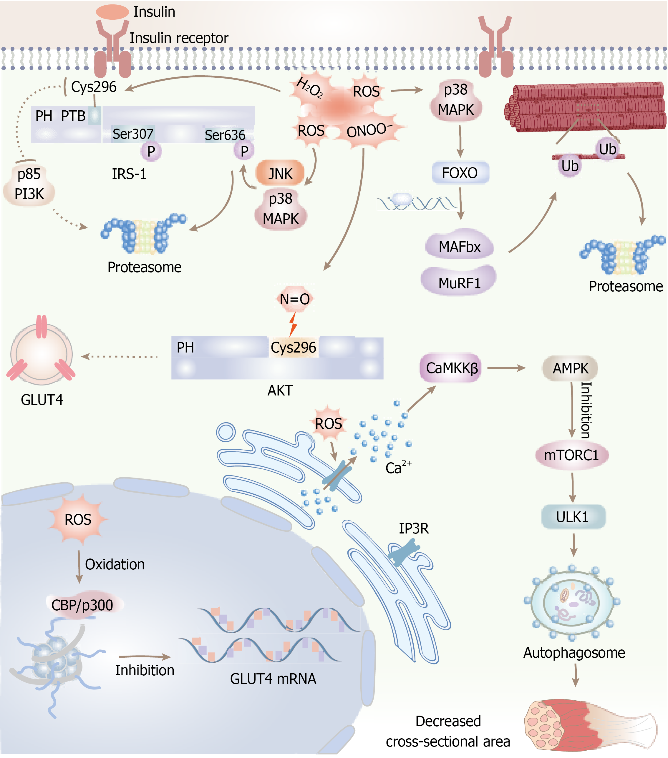
Figure 3 Reactive oxygen species contribute to insulin resistance and muscle atrophy in skeletal muscle through several molecular mechanisms.
Specifically, hydrogen peroxide (H2O2) directly oxidizes cysteine residue 296 on insulin receptor substrate-1, thereby inhibiting the recruitment of phosphoinositide 3-kinase. Reactive oxygen species (ROS)-mediated activation of JNK and p38 MAPK leads to the inhibitory phosphorylation of insulin receptor substrate-1 at serine residues 307 and 636, which facilitates its ubiquitination and subsequent proteasomal degradation. Peroxynitrite (ONOO-) nitrates cysteine 296 on protein kinase B, resulting in diminished kinase activity and impaired translocation of GLUT4. Furthermore, ROS induces oxidation of the transcriptional coactivators CBP/p300, causing epigenetic repression of GLUT4 gene expression. Concurrently, ROS activates the p38 MAPK- forkhead box O transcription factors signaling axis, which upregulates the expression of muscle-specific E3 ubiquitin ligases MAFbx and MuRF1, thereby promoting proteasomal protein degradation. Moreover, ROS triggers calcium release through oxidation of IP3R, which activates the Ca2+/CaMKKβ/AMP-AMP-activated protein kinase pathway. This activation inhibits mechanistic target of rapamycin complex 1 and stimulates ULK1-mediated autophagy, collectively contributing to a reduction in muscle fiber cross-sectional area. ROS: Reactive oxygen species; IRS-1: Insulin receptor substrate-1; PI3K: Phosphoinositide 3-kinase; AKT: Protein kinase B; FOXO: Forkhead box O transcription factors; mTORC1: Mechanistic target of rapamycin complex 1.
- Citation: Xie XL, Cao HY, Wang ZT, Chen HR, Zhou LS, Qian T, Han ZY. Pathophysiological mechanisms and risk factors linking type 2 diabetes mellitus and sarcopenia: A narrative review. World J Diabetes 2026; 17(10): 124847
- URL: https://www.wjgnet.com/1948-9358/full/v17/i10/124847.htm
- DOI: https://dx.doi.org/10.4239/wjd.124847