
















Copyright: ©Author(s) 2026.
World J Diabetes. Oct 15, 2026; 17(10): 124847
Published online Oct 15, 2026. doi: 10.4239/wjd.124847
Published online Oct 15, 2026. doi: 10.4239/wjd.124847
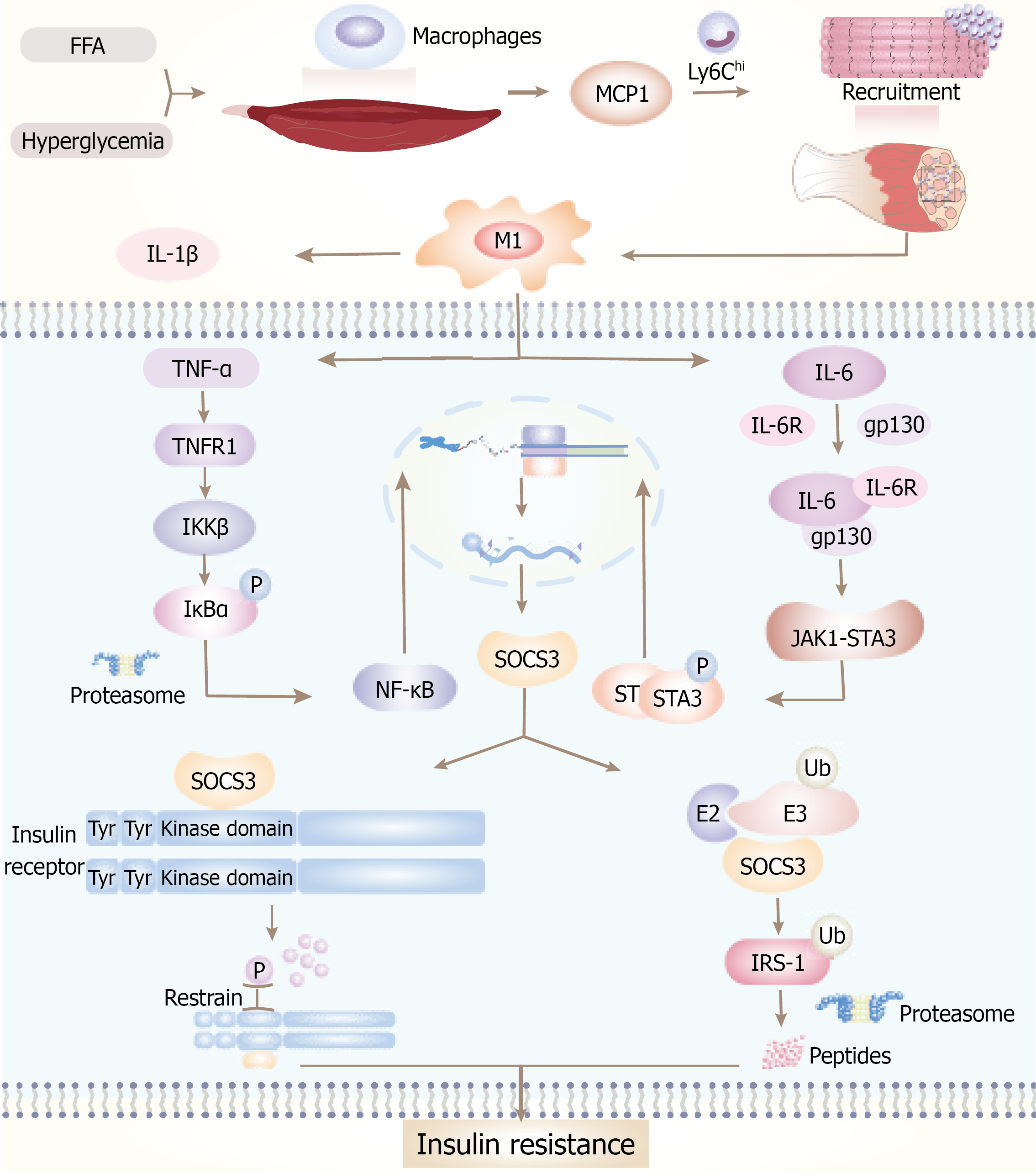
Figure 2 Inflammation induces insulin resistance in skeletal muscle.
Elevated levels of glucose and free fatty acid stimulate the secretion of monocyte chemoattractant protein-1 from both muscle fibers and resident macrophages. This chemokine facilitates the recruitment of Ly6Chi monocytes, which subsequently differentiate into pro-inflammatory M1 macrophages. These macrophages secrete pro-inflammatory cytokines, including tumor necrosis factor-alpha, interleukin (IL)-6, and IL-1β. tumor necrosis factor-alpha activates the IKKβ-IκBα-nuclear factor kappa-B signaling cascade, whereas IL-6 induces phosphorylation of the JAK1-STAT3 pathway. The convergence of these signaling pathways leads to the upregulation of suppressor of cytokine signaling 3, which impairs insulin signaling by inhibiting insulin receptor autophosphorylation and promoting the ubiquitination and subsequent degradation of insulin receptor substrate-1. Collectively, these molecular events culminate in the development of insulin resistance within skeletal muscle tissue. MCP-1: Monocyte chemoattractant protein-1; TNF-α: Tumor necrosis factor-alpha; IL-6: Interleukin-6; SOCS3: Suppressor of cytokine signaling 3; FFA: Free fatty acid; IRS-1: Insulin receptor substrate-1.
- Citation: Xie XL, Cao HY, Wang ZT, Chen HR, Zhou LS, Qian T, Han ZY. Pathophysiological mechanisms and risk factors linking type 2 diabetes mellitus and sarcopenia: A narrative review. World J Diabetes 2026; 17(10): 124847
- URL: https://www.wjgnet.com/1948-9358/full/v17/i10/124847.htm
- DOI: https://dx.doi.org/10.4239/wjd.124847