
















Copyright: ©Author(s) 2026.
World J Diabetes. Oct 15, 2026; 17(10): 124847
Published online Oct 15, 2026. doi: 10.4239/wjd.124847
Published online Oct 15, 2026. doi: 10.4239/wjd.124847
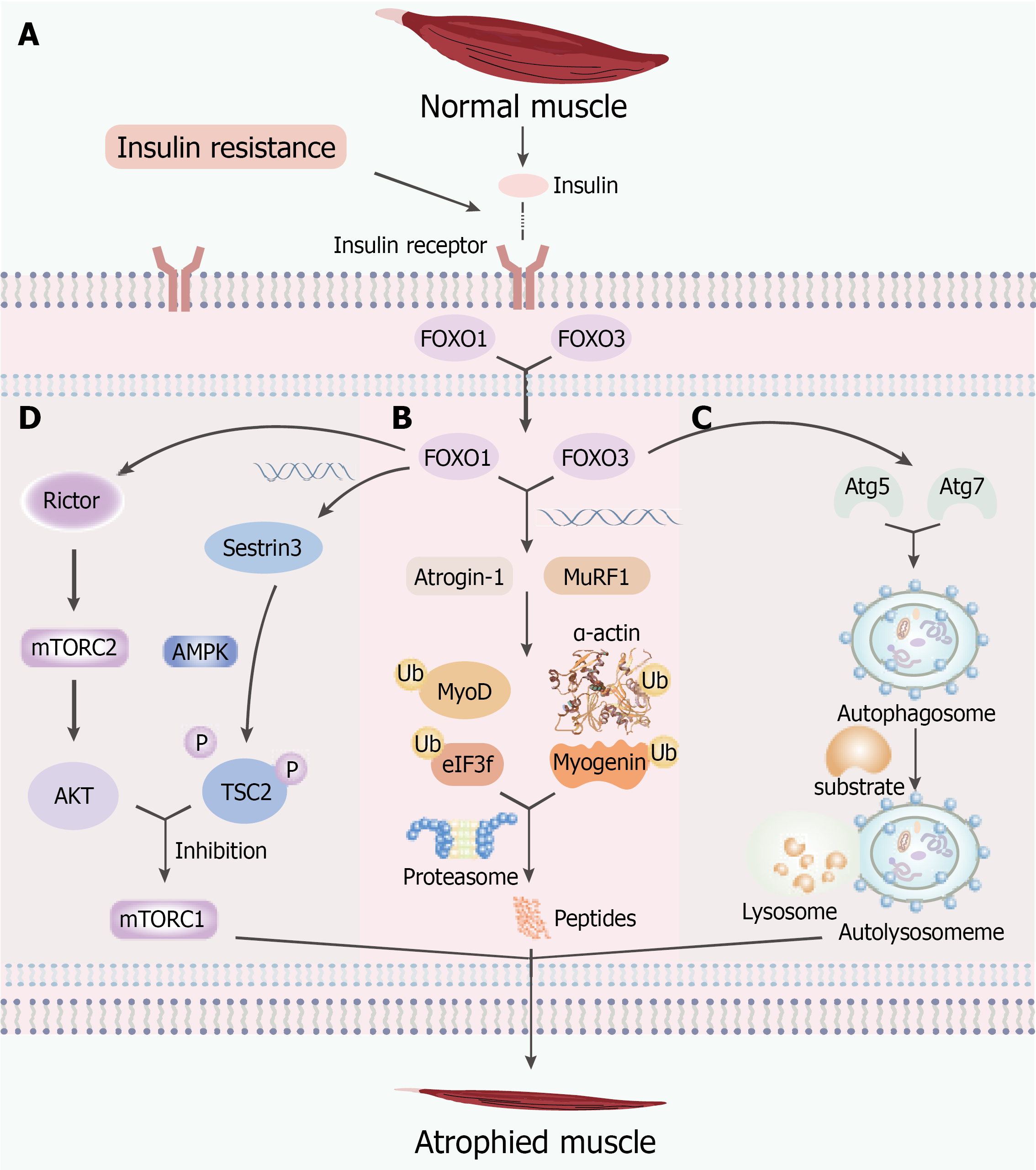
Figure 1 Mechanisms of insulin resistance-induced skeletal muscle atrophy.
A: Insulin resistance impairs insulin receptor-mediated phosphoinositide 3-kinase-protein kinase B (AKT) signaling, resulting in reduced AKT activity and subsequent nuclear translocation of forkhead box O transcription factors (FOXO) 1 and FOXO3; B: Activated FOXO1/FOXO3 promote the transcription of muscle-specific ubiquitin ligases, including Atrogin-1 and MuRF1, which induce ubiquitination and proteasomal degradation of myogenic regulatory proteins (MyoD, myogenin, and eIF3f) and contractile proteins (α-actin), thereby accelerating muscle protein degradation; C: FOXO3 activates autophagy-related genes, including Atg5 and Atg7, promoting autophagosome formation and autolysosomal degradation, which further contributes to skeletal muscle wasting; D: FOXO1 inhibits mechanistic target of rapamycin complex 1-mediated protein synthesis through both tuberous sclerosis complex 2 (TSC2)-dependent and TSC2-independent pathways. The Sestrin3-AMP-activated protein kinase-TSC2 pathway and rapamycin-insensitive companion of mechanistic target of rapamycin-mechanistic target of rapamycin complex 2-AKT pathway suppress mechanistic target of rapamycin complex 1 activity, leading to impaired muscle protein synthesis. AKT: Protein kinase B; FOXO: Forkhead box O transcription factors; mTORC1: Mechanistic target of rapamycin complex 1; mTORC2: Mechanistic target of rapamycin complex 2; TSC2: Tuberous sclerosis complex 2; Rictor: Rapamycin-insensitive companion of mechanistic target of rapamycin; AMPK: AMP-activated protein kinase.
- Citation: Xie XL, Cao HY, Wang ZT, Chen HR, Zhou LS, Qian T, Han ZY. Pathophysiological mechanisms and risk factors linking type 2 diabetes mellitus and sarcopenia: A narrative review. World J Diabetes 2026; 17(10): 124847
- URL: https://www.wjgnet.com/1948-9358/full/v17/i10/124847.htm
- DOI: https://dx.doi.org/10.4239/wjd.124847