
















©The Author(s) 2026.
World J Diabetes. Jan 15, 2026; 17(1): 112027
Published online Jan 15, 2026. doi: 10.4239/wjd.v17.i1.112027
Published online Jan 15, 2026. doi: 10.4239/wjd.v17.i1.112027
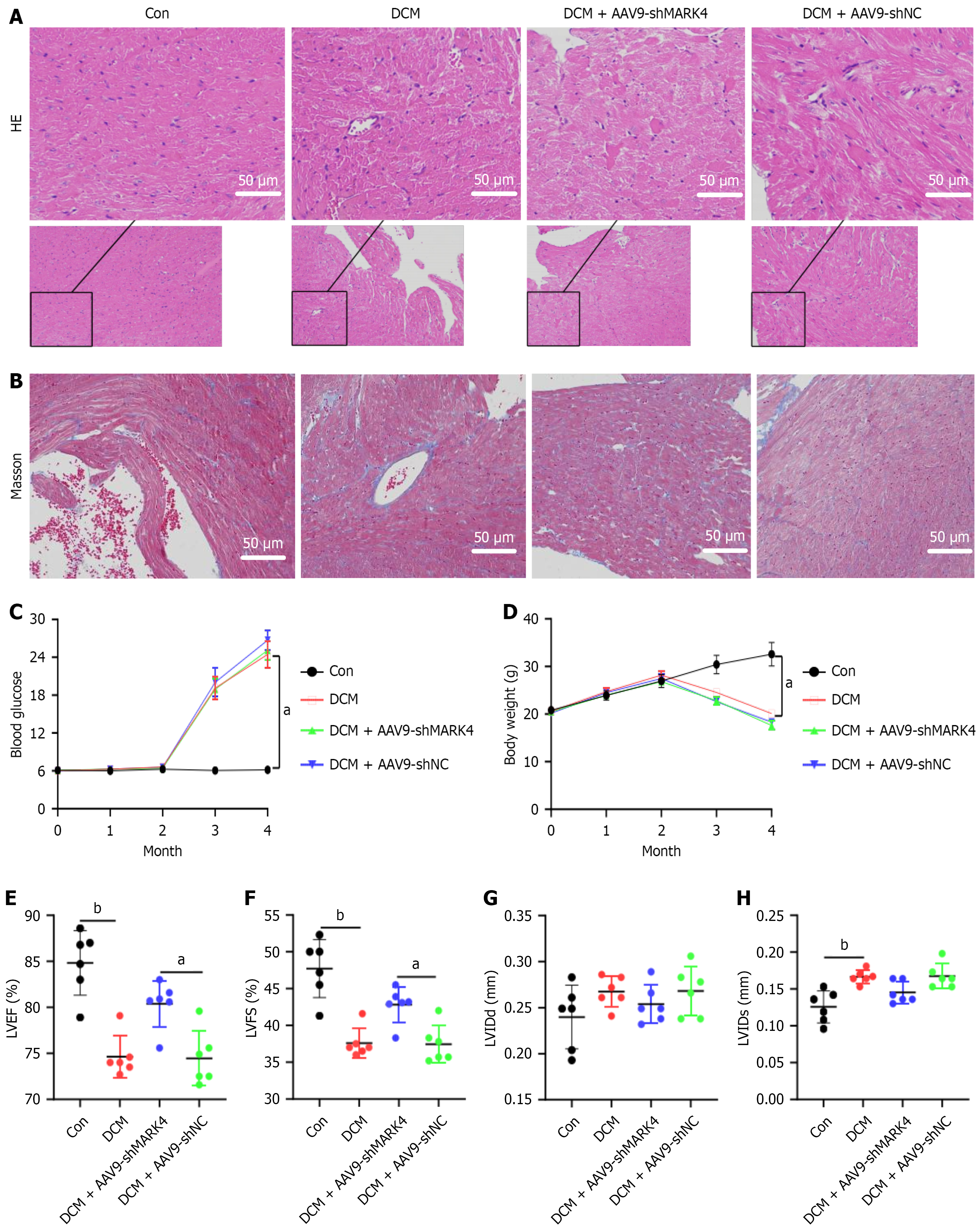
Figure 3 Impact of microtubule affinity-regulating kinase 4 knockdown on myocardial injury and fibrosis, blood glucose, body weight, and cardiac function and structure in diabetic cardiomyopathy model mice.
A: Hematoxylin and eosin staining of left ventricular tissue (× 200); B: Masson’s staining of left ventricular tissue (× 200); C: Blood glucose levels; D: Body weights; E: The left ventricular ejection fractions; F: The left ventricular short axis shortening rates; G: Left ventricular end-diastolic diameters; H: Left ventricular end-systolic diameters. Values are mean ± SD (n = 4). aP < 0.05, and bP < 0.01. HE: Hematoxylin and eosin; Con: Control; DCM: Diabetic cardiomyopathy; MARK4: Microtubule affinity-regulating kinase 4; sh: Small hairpin; NC: Negative control; LVEF: Left ventricular ejection fractions; LVFS: Left ventricular shortening fraction; LVIDd: Left ventricular end-diastolic diameters; LVIDs: Left ventricular end-systolic diameter; AAV9: Adeno-associated virus 9.
- Citation: Wu Y, Wang WY, Zhang JQ, Wang S, Zeng Z, Fu L, Li B. Microtubule affinity-regulating kinase 4 exacerbates diabetic cardiomyopathy by inhibiting UNC-51-like kinase 1-mediated mitochondrial autophagy. World J Diabetes 2026; 17(1): 112027
- URL: https://www.wjgnet.com/1948-9358/full/v17/i1/112027.htm
- DOI: https://dx.doi.org/10.4239/wjd.v17.i1.112027