
















©The Author(s) 2026.
World J Diabetes. Jan 15, 2026; 17(1): 111165
Published online Jan 15, 2026. doi: 10.4239/wjd.v17.i1.111165
Published online Jan 15, 2026. doi: 10.4239/wjd.v17.i1.111165
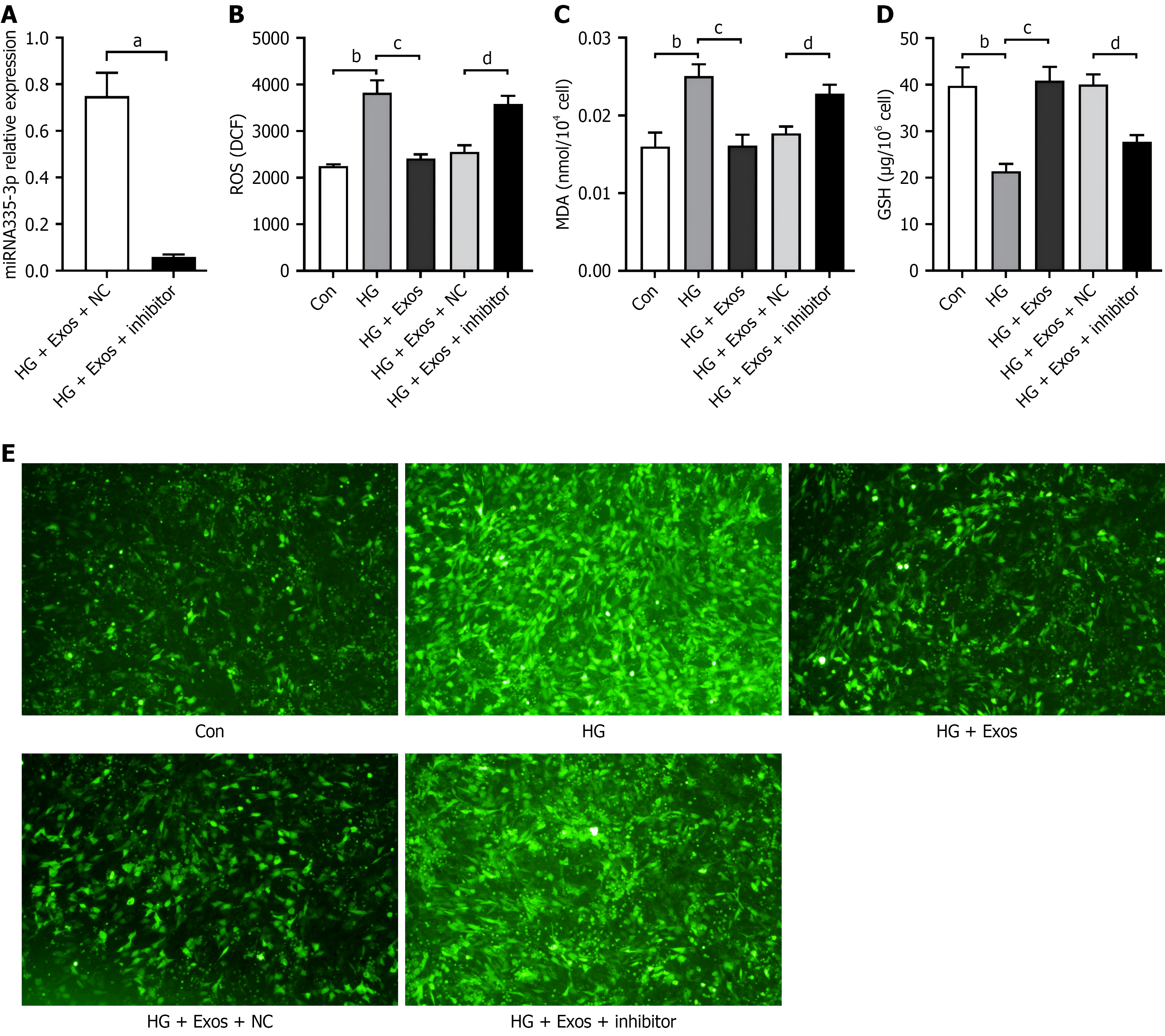
Figure 5 Inhibition of microRNA-335-3p abolishes endothelial cell-exosomes-mediated protection against high glucose-induced oxidative stress in osteoblasts.
A: Detection of microRNA-335-3p levels in osteoblasts induced by high glucose after inhibition of microRNA-335-3p by reverse trans
- Citation: Shao C, Zhang LJ, Song YL, Wang YQ, Zha XJ, Li J, Ye CS, Chen LL, Chen MW, Jin GX. Endothelial cell-derived exosomes inhibit high glucose-induced osteoblast ferroptosis by activating microRNA-335-3p/prostaglandin endoperoxide synthase 2. World J Diabetes 2026; 17(1): 111165
- URL: https://www.wjgnet.com/1948-9358/full/v17/i1/111165.htm
- DOI: https://dx.doi.org/10.4239/wjd.v17.i1.111165