
















©The Author(s) 2025.
World J Diabetes. Aug 15, 2025; 16(8): 106833
Published online Aug 15, 2025. doi: 10.4239/wjd.v16.i8.106833
Published online Aug 15, 2025. doi: 10.4239/wjd.v16.i8.106833
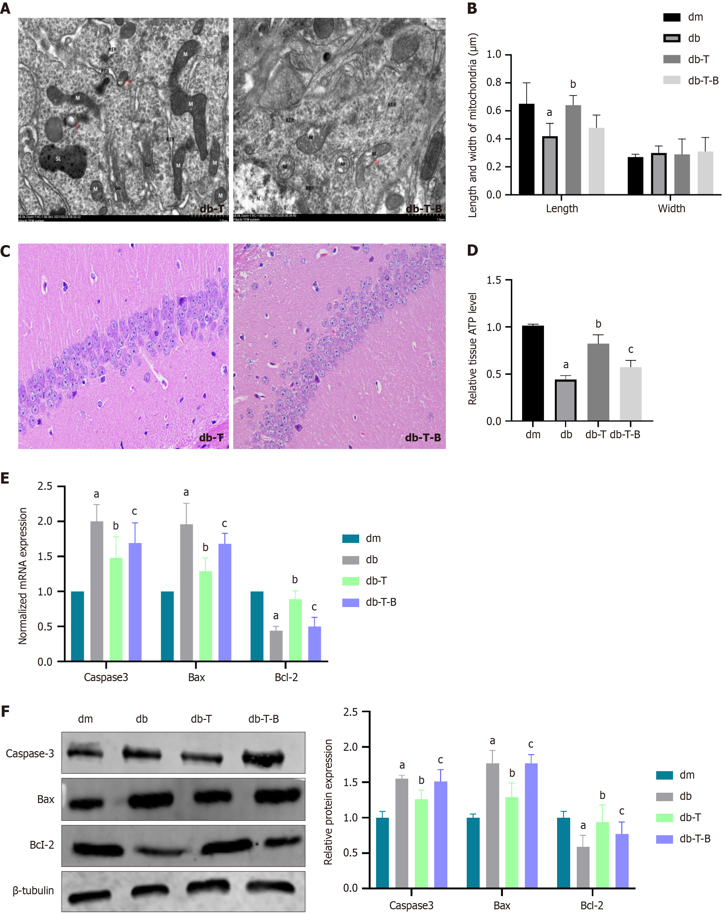
Figure 9 Troxerutin improved mitochondrial fission and cell apoptosis in the hippocampal tissue of diabetic cognitive dysfunction mice.
A: Transmission electron microscopy images show a decrease in mitochondrial fission in the hippocampal tissue of db-T group compared to db group; B: The length and width of mitochondria were measured (n = 20); C: Hematoxylin and eosin staining shows that hippocampal pyramidal cells injury was improved in db-T group compared to db group (400 ×); D: Relative of adenosine triphosphate level; E: The normalized mRNA expression of caspase-3, B-cell lymphoma 2 (Bcl-2)-associated X protein (Bax), and Bcl-2; F: The relative protein expression of The relative protein expression of caspase-3, Bcl-2-associated X protein, and Bcl-2. Bradykinin is a transient receptor potential melastatin 7 agonist, and its addition diminished the neuroprotective effect of troxerutin. aP < 0.05, compared with dm group; bP < 0.05, compared with diabetes group; cP < 0.05, compared with diabetes + troxerutin group. Data presented as mean ± SEM. dm: Control group; dm-T: Control + troxerutin group; db: Diabetes group; db-T: Diabetes + troxerutin group; db-T-B: Diabetes + troxerutin + bradykinin group; Bax: B-cell lymphoma 2-associated X protein; Bcl-2: B-cell lymphoma 2.
- Citation: Li J, Gao M, Wang JX, Li HY, Wang P, Yuan F, Liu AJ, Zhang SY. Troxerutin improves diabetic cognitive dysfunction by inhibiting mitochondrial fission mediated by transient receptor potential melastatin 7/calcineurin/dynamin-related protein 1ser637. World J Diabetes 2025; 16(8): 106833
- URL: https://www.wjgnet.com/1948-9358/full/v16/i8/106833.htm
- DOI: https://dx.doi.org/10.4239/wjd.v16.i8.106833