











BPG is committed to discovery and dissemination of knowledge
Review
©The Author(s) 2025.
World J Diabetes. Dec 15, 2025; 16(12): 114395
Published online Dec 15, 2025. doi: 10.4239/wjd.v16.i12.114395
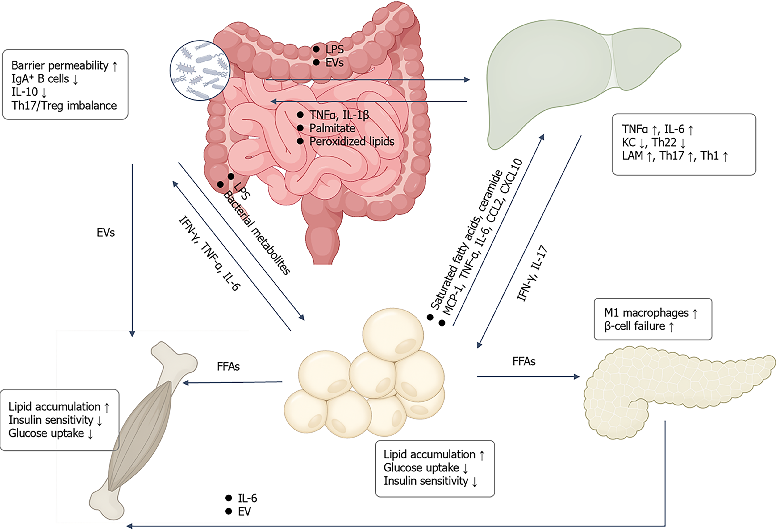
Figure 4
Figure 4 Inter-organ crosstalk drives metabolic inflammation and insulin resistance. Intestinal dysbiosis and barrier impairment allow lipopolysaccharide and extracellular vesicles (EVs) to enter the liver via the portal vein, triggering hepatic inflammation [tumor necrosis factor-α, interleukin (IL)-1β] and synergizing with lipotoxic mediators such as palmitic acid and oxidized lipids to amplify inflammatory responses. Liver-derived inflammatory cytokines and helper T (Th) 1/Th17 skewing further exacerbate intestinal mucosal immune dysregulation. Under lipid overload, adipose tissue releases free fatty acids, ceramides, and chemokines/cytokines including monocyte chemoattractant protein-1, IL-6, CCL2, and CXCL10, promoting hepatic immune remodeling and inflammation (reduced Kupffer cells, increased triggering receptor expressed on myeloid cells 2+ lipid-associated macrophages, Th1/Th17 polarization, and Th22 suppression). Adipose-derived free fatty acids concurrently contribute to lipid deposition in skeletal muscle, reduced insulin sensitivity, and impaired glucose uptake, while also inducing M1 macrophage accumulation and β-cell dysfunction in the pancreas. Skeletal muscle-derived IL-6 and inflammatory EVs can further circulate to the liver, aggravating hepatic insulin resistance. These interactions form a positive feedback loop that collectively drives the progression of systemic insulin resistance and hyperglycemia. IL: Interleukin; IgA: Immunoglobulin A; Treg: Regulatory T cell; Th: Helper T; EV: Extracellular vesicle; TNF: Tumor necrosis factor; LPS: Lipopolysaccharide; IFN: Interferon; FFAs: Free fatty acids; LAM: Lipid-associated macrophage; KC: Kupffer cell; MCP-1: Monocyte chemoattractant protein-1.


Write to the Help Desk