
















©The Author(s) 2025.
World J Diabetes. Dec 15, 2025; 16(12): 114395
Published online Dec 15, 2025. doi: 10.4239/wjd.v16.i12.114395
Published online Dec 15, 2025. doi: 10.4239/wjd.v16.i12.114395
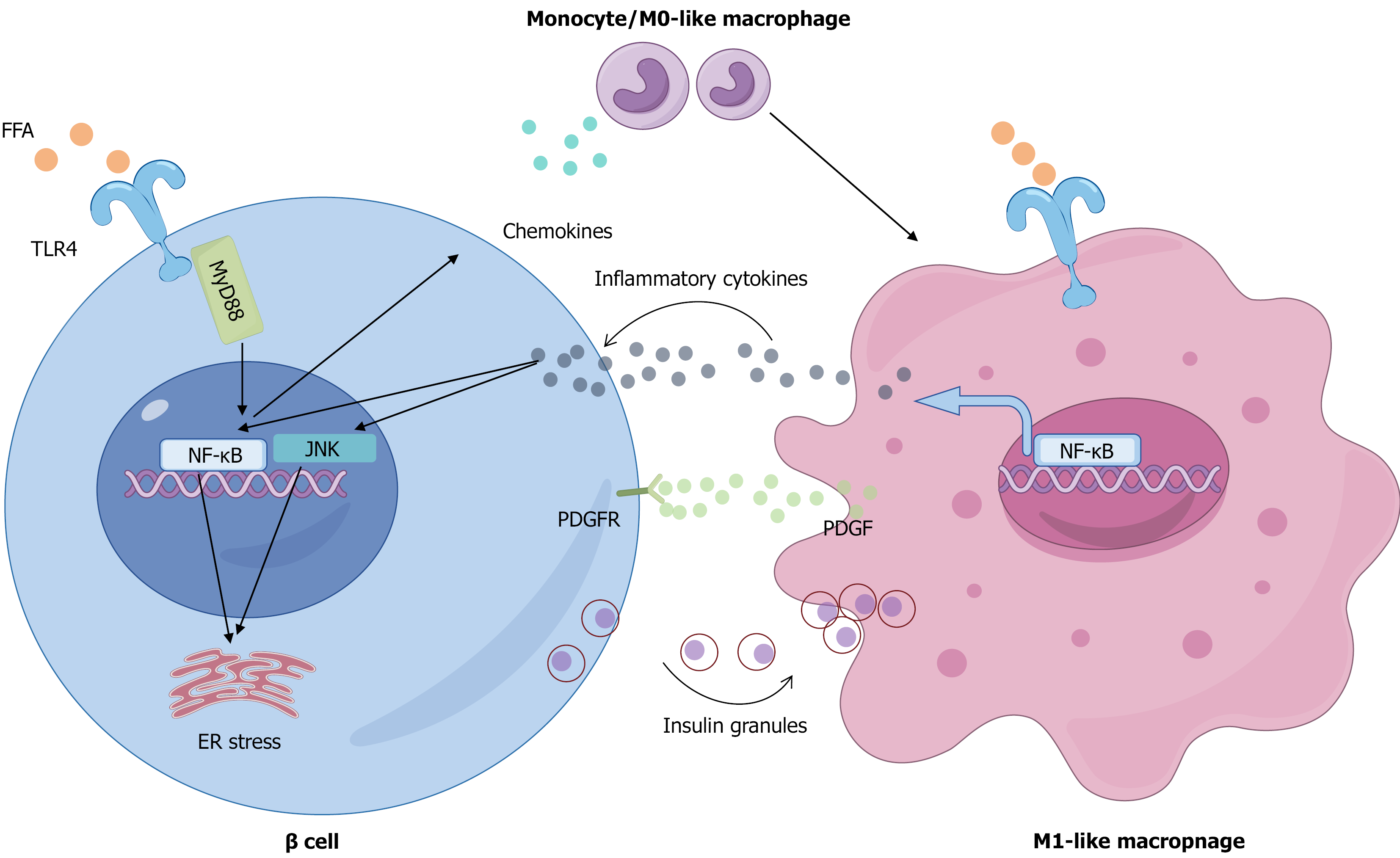
Figure 2 Mechanisms of macrophage-mediated islet inflammation and β-cell dysfunction induced by disordered lipid metabolism.
Elevated circulating free fatty acids initiate islet inflammation by activating the Toll-like receptor (TLR) 4/nuclear factor kappa-B (NF-κB) pathway in pancreatic β-cells, prompting them to secrete chemokines that recruit circulating monocytes. Upon infiltration and under lipotoxic conditions, these monocytes polarize into pro-inflammatory M1 macrophages, which are further activated via the TLR4/NF-κB pathway to secrete abundant inflammatory cytokines. These cytokines in turn impair β-cell function by activating intracellular Janus tyrosine kinase and NF-κB signaling and inducing endoplasmic reticulum stress, collectively suppressing glucose-stimulated insulin secretion and insulin gene expression. Additionally, the activated M1 macrophages directly phagocytose insulin secretory granules and release platelet-derived growth factor to engage platelet-derived growth factor receptors on β-cells, stimulating a compensatory proliferative response that ultimately fails to prevent β-cell failure and inadequate insulin secretion. FFAs: Free fatty acids; TLR: Toll-like receptor; NF-κB: Nuclear factor kappa-B; JNK: Janus tyrosine kinase; ER: Endoplasmic reticulum; MyD88: Myeloid differentiation primary response 88; PDGF: Platelet-derived growth factor; PDGFR: Platelet-derived growth factor receptor.
- Citation: Yang HY, Wei Y, Mao Q, Zhao LH. Immune activation induced by dysregulated lipid metabolism in the pathogenesis of type 2 diabetes. World J Diabetes 2025; 16(12): 114395
- URL: https://www.wjgnet.com/1948-9358/full/v16/i12/114395.htm
- DOI: https://dx.doi.org/10.4239/wjd.v16.i12.114395