
















©The Author(s) 2025.
World J Diabetes. Nov 15, 2025; 16(11): 109859
Published online Nov 15, 2025. doi: 10.4239/wjd.v16.i11.109859
Published online Nov 15, 2025. doi: 10.4239/wjd.v16.i11.109859
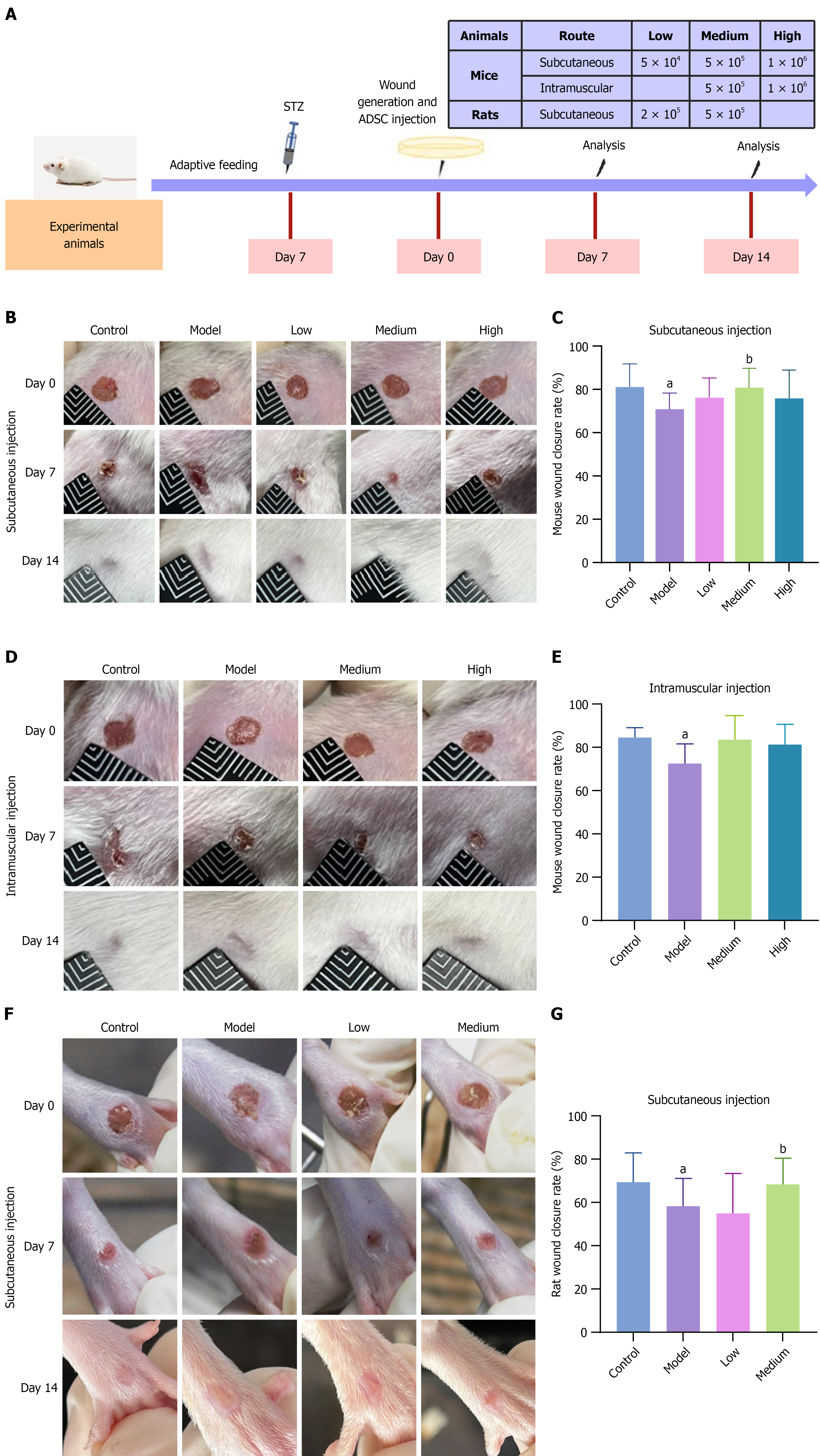
Figure 2 Wound closure in diabetic foot ulcer model mice and rats after treatment with adipose-derived mesenchymal stem cells.
A: Schematic diagram of the animal experiment; B: Representative images of ulcers in diabetic foot ulcer (DFU) mice on days 7 and 14 after subcutaneous injection of low, medium, or high doses of adipose-derived mesenchymal stem cells (ADSCs) or vehicle; C: Statistical bar chart of the wound closure rate in DFU mice on day 7 after subcutaneous injection. The wound healing rate in the medium-dose group was significantly greater than that in the model group (P < 0.05), n = 14; D: Representative images of ulcers in DFU mice on days 7 and 14 after intramuscular injection of medium and high ADSC doses; E: Statistical chart of the wound closure rate in DFU mice on day 7 after intramuscular injection, n = 7; F: Representative images of ulcers in DFU rats on days 7 and 14 after subcutaneous injection of low and medium ADSC doses; G: Statistical chart of the wound closure rate in DFU rats on day 7 after subcutaneous injection. The wound healing rate in the medium-dose group was significantly greater than that in the model group (P < 0.05), n = 14. The data are presented as mean ± SD. STZ: Streptozotocin; ADSC: Adipose-derived mesenchymal stem cell. aP < 0.05 vs control, and bP < 0.05 vs model.
- Citation: Cao J, Liu ZC, An WQ, Zhang S, Zhang X, Li LJ, Ji HL, Long X, Yang YM. Optimizing adipose-derived stem cell therapy for diabetic foot ulcers. World J Diabetes 2025; 16(11): 109859
- URL: https://www.wjgnet.com/1948-9358/full/v16/i11/109859.htm
- DOI: https://dx.doi.org/10.4239/wjd.v16.i11.109859