
















©The Author(s) 2025.
World J Diabetes. Nov 15, 2025; 16(11): 109859
Published online Nov 15, 2025. doi: 10.4239/wjd.v16.i11.109859
Published online Nov 15, 2025. doi: 10.4239/wjd.v16.i11.109859
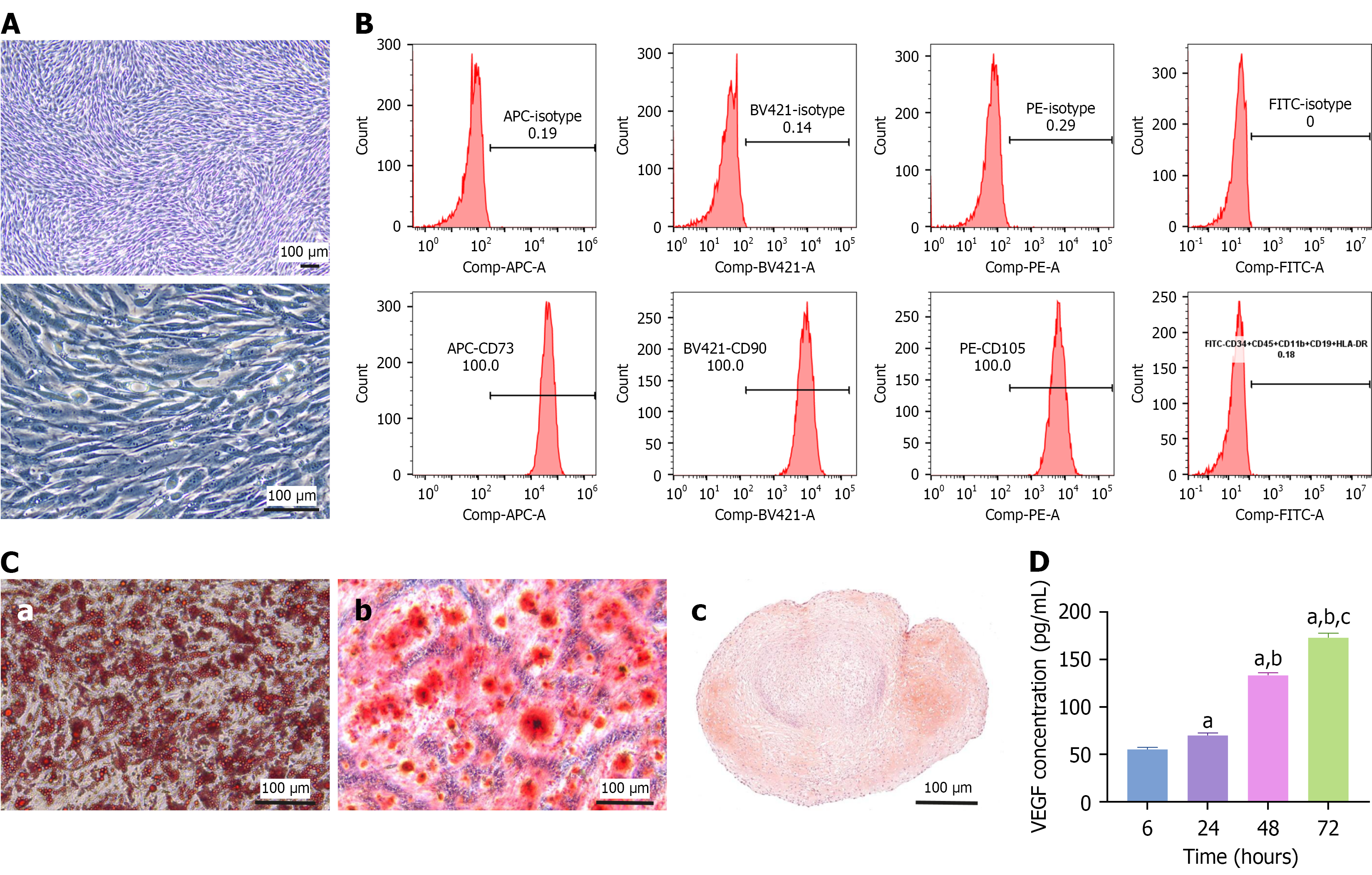
Figure 1 Characterization of adipose-derived mesenchymal stem cells.
A: Representative cell morphology images of adipose-derived mesenchymal stem cells (ADSCs) at 5 × and 20 ×; B: Flow cytometry indicated that CD73, CD105, and CD90 were highly expressed and that CD11b, CD19, CD34, CD45, and human leukocyte antigen-DR were negligibly expressed by ADSCs; C: Triplet differentiation assays revealed that ADSCs could differentiate into adipocytes (a), osteocytes (b), and chondrocytes (c), which were stained with Oil Red O, alizarin red, and safranin O, respectively; D: Enzyme-linked immunosorbent assay was used to measure the concentration of vascular endothelial growth factor secreted by ADSCs cultured for 6, 24, 48, and 72 hours. The data are presented as mean ± SD. Scale bar, 100 μm. Comp: Compensation; APC: Allophycocyanin; A: Area; BV421: Brilliant violet 421; PE: Phycoerythrin; FITC: Fluorescein isothiocyanate; HLA-DR: Human leukocyte antigen-DR; VEGF: Vascular endothelial growth factor. aP < 0.0001 vs 6 hours, bP < 0.0001 vs 24 hours, and cP < 0.0001 vs 48 hours.
- Citation: Cao J, Liu ZC, An WQ, Zhang S, Zhang X, Li LJ, Ji HL, Long X, Yang YM. Optimizing adipose-derived stem cell therapy for diabetic foot ulcers. World J Diabetes 2025; 16(11): 109859
- URL: https://www.wjgnet.com/1948-9358/full/v16/i11/109859.htm
- DOI: https://dx.doi.org/10.4239/wjd.v16.i11.109859