
















©The Author(s) 2025.
World J Diabetes. Oct 15, 2025; 16(10): 110631
Published online Oct 15, 2025. doi: 10.4239/wjd.v16.i10.110631
Published online Oct 15, 2025. doi: 10.4239/wjd.v16.i10.110631
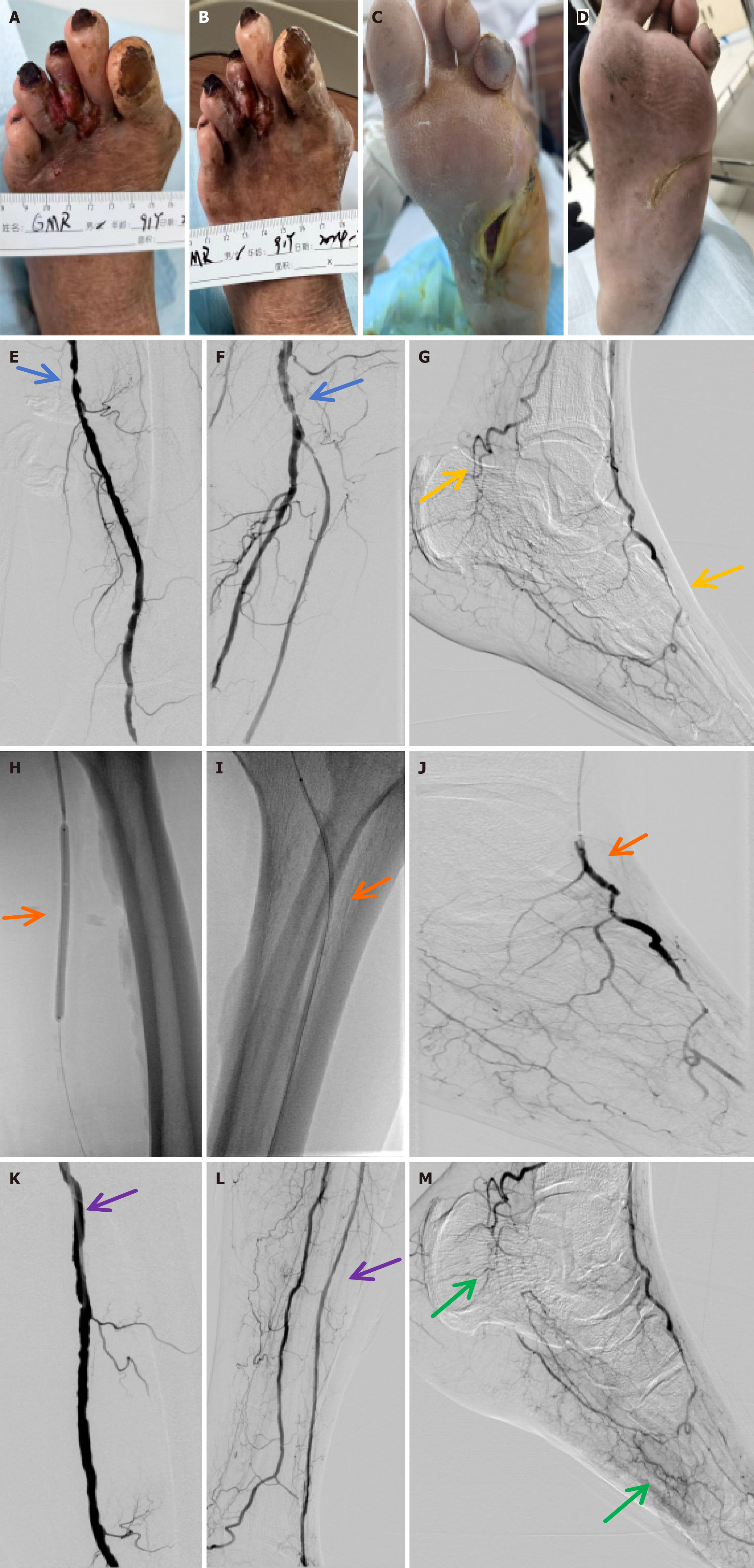
Figure 6 Diabetic foot ulcer repair.
Diabetic foot ulcers of patients in each group before and after treatment (blue arrows indicate lower extremity vascular stenosis, yellow arrows indicate lack of microcirculation in the foot, orange arrows indicate balloon dilatation and opening of blood vessels, purple arrows indicate revascularization of lower extremity vessels, and green arrows indicate establishment of microcirculation in the foot). A and B: Platelet-rich plasma (PRP) + endovascular angioplasty group before treatment (A), and PRP + endovascular angioplasty 1 month after treatment (B); C and D: PRP + endovascular angioplasty group before treatment (C), and PRP + endovascular angioplasty 1 month after treatment (D); E-G: PRP + endovascular angioplasty group before endovascular angioplasty under digital subtraction angiography (DSA); H-J: PRP + endovascular angioplasty group during endovascular angioplasty under DSA; K-M: PRP + endovascular angioplasty group 1 month after endovascular angioplasty under DSA.
- Citation: Huang C, Liu HZ, Liang JB, Zhao W, Wang YS, Ruan LF, Zhuang WZ, Li YS, Wang Q, Tang YK. In vitro and clinical evaluation of platelet-rich plasma combined with angioplasty in diabetic foot treatment. World J Diabetes 2025; 16(10): 110631
- URL: https://www.wjgnet.com/1948-9358/full/v16/i10/110631.htm
- DOI: https://dx.doi.org/10.4239/wjd.v16.i10.110631