
















©The Author(s) 2025.
World J Diabetes. Oct 15, 2025; 16(10): 110041
Published online Oct 15, 2025. doi: 10.4239/wjd.v16.i10.110041
Published online Oct 15, 2025. doi: 10.4239/wjd.v16.i10.110041
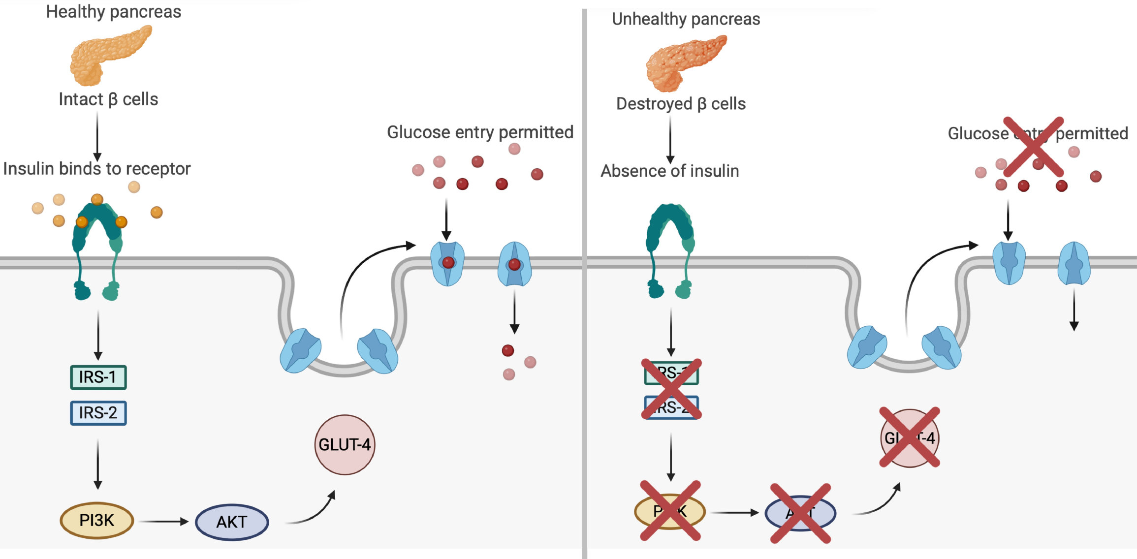
Figure 5 Insulin signaling cascade and glucose transporter type 4 translocation in type 1 diabetes.
This figure illustrates the effects of healthy vs impaired pancreatic function on glucose metabolism in type 1 diabetes. In the healthy pancreas (panel A, left), functional β cells secrete insulin, which binds to its receptor on the cell membrane and activates a signaling cascade via insulin receptor substrate 1 (IRS-1) and IRS-2. This triggers the phosphoinositide 3-kinase (PI3K)/protein kinase B (PKB) pathway, promoting the translocation of glucose transporter type 4 (GLUT4) to the membrane and facilitating glucose entry into muscle cells. By contrast, panel B (right) depicts the absence of insulin secretion due to β-cell destruction, which disrupts activation of the IRS-1/IRS-2 and PI3K/PKB pathways. As a result, GLUT4 translocation is impaired, preventing glucose uptake and leading to hyperglycemia. This dysfunction is closely associated with inflammatory and oxidative stress in type 1 diabetes, reinforcing the importance of therapeutic strategies that preserve β-cell function or modulate inflammation.
- Citation: Ramos Fernandes VA, Mendes LR, Franco Netto ROR, Belozo FL, Bezerra AA, dos Santos CPC, Cruel PTE, Buchaim DV, Buchaim RL, da Cunha MR. Anti-inflammatory effects of cannabidiol in the treatment of type 1 diabetes: A mini review. World J Diabetes 2025; 16(10): 110041
- URL: https://www.wjgnet.com/1948-9358/full/v16/i10/110041.htm
- DOI: https://dx.doi.org/10.4239/wjd.v16.i10.110041