
















©The Author(s) 2025.
World J Diabetes. Oct 15, 2025; 16(10): 109815
Published online Oct 15, 2025. doi: 10.4239/wjd.v16.i10.109815
Published online Oct 15, 2025. doi: 10.4239/wjd.v16.i10.109815
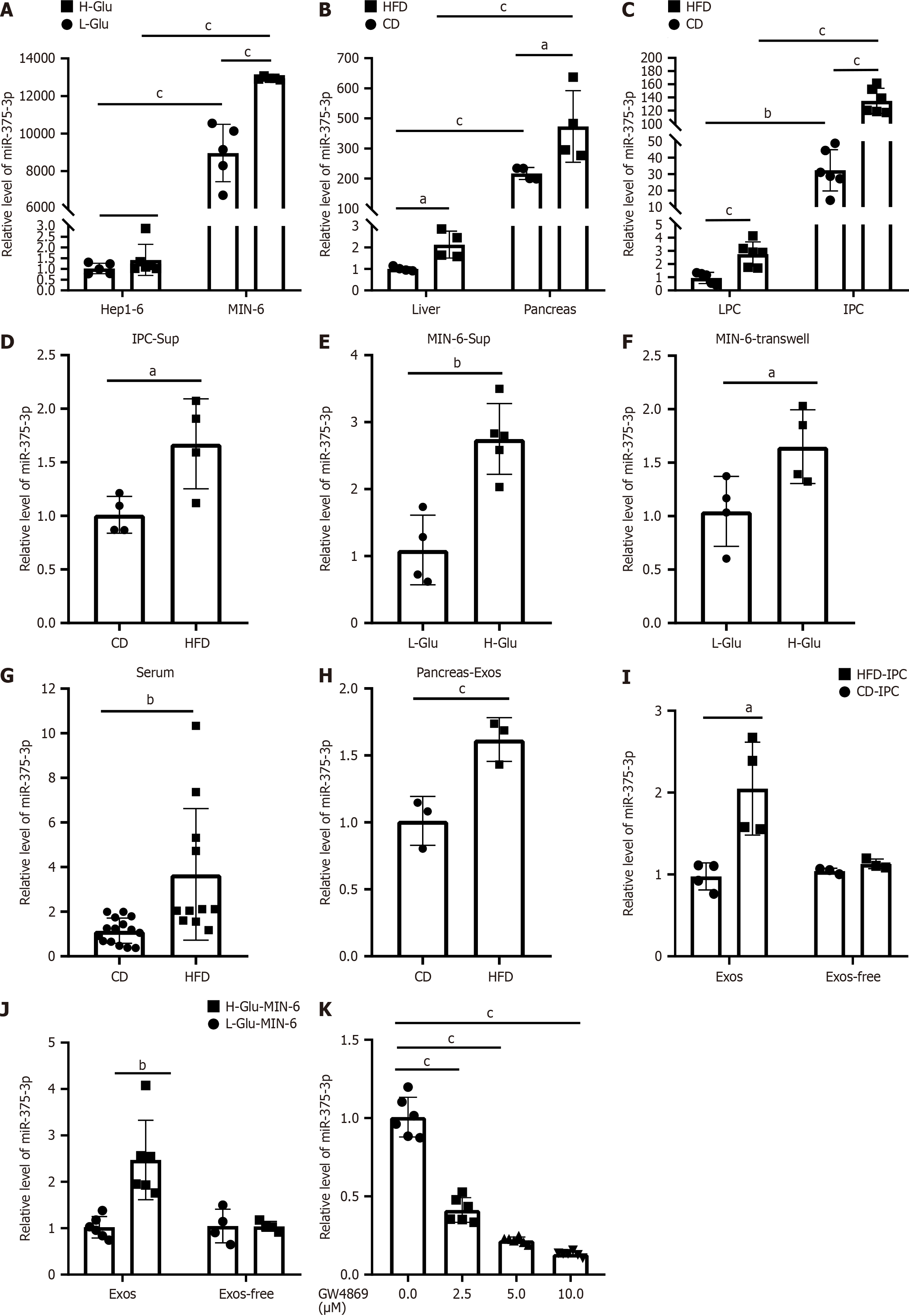
Figure 4 miR-375-3p is transferred from glucotoxic pancreatic cells to hepatocytes via exosomes.
Relative miR-375-3p levels were quantified by quantitative reverse transcription polymerase chain reaction. A: Hep1-6 and MIN-6 cells after low- or high-glucose stimulation; B: Pancreatic and liver tissues of high-fat diet (HFD)-fed or chow diet (CD)-fed mice; C: Primary islet cells (IPCs) and LPCs from HFD-fed or CD-fed mice; D: Hep1-6 cells treated with conditioned media from IPCs (IPC-Sup); E: Hep1-6 cells treated with conditioned media from MIN-6 cells (MIN6-Sup); F: Hep1-6 cells cocultured with MIN-6 cells using Transwell plates (MIN6-Transwell); G: Plasma of HFD-fed or CD-fed mice; H-J: Exosomes or exosome-depleted supernatantsisolated from pancreatic tissues (pancreas-Exos), IPCs (IPC-Exos), and MIN-6 cells (MIN-6 Exos); K: Hep1-6 cells treatment with conditioned media from GW4869-treated MIN-6 cells. The experiments were repeated three times. aP < 0.05, bP < 0.01, cP < 0.001.
- Citation: Xu FZ, Dou L, Wu X, Xia CX, Yu DN, Man Y, Shen T, Huang XQ. Exosomal transfer of miR-375-3p from pancreatic β cells to hepatocytes impairs hepatic glycogenesis via Rbpj repression. World J Diabetes 2025; 16(10): 109815
- URL: https://www.wjgnet.com/1948-9358/full/v16/i10/109815.htm
- DOI: https://dx.doi.org/10.4239/wjd.v16.i10.109815