
















©The Author(s) 2025.
World J Diabetes. Oct 15, 2025; 16(10): 109782
Published online Oct 15, 2025. doi: 10.4239/wjd.v16.i10.109782
Published online Oct 15, 2025. doi: 10.4239/wjd.v16.i10.109782
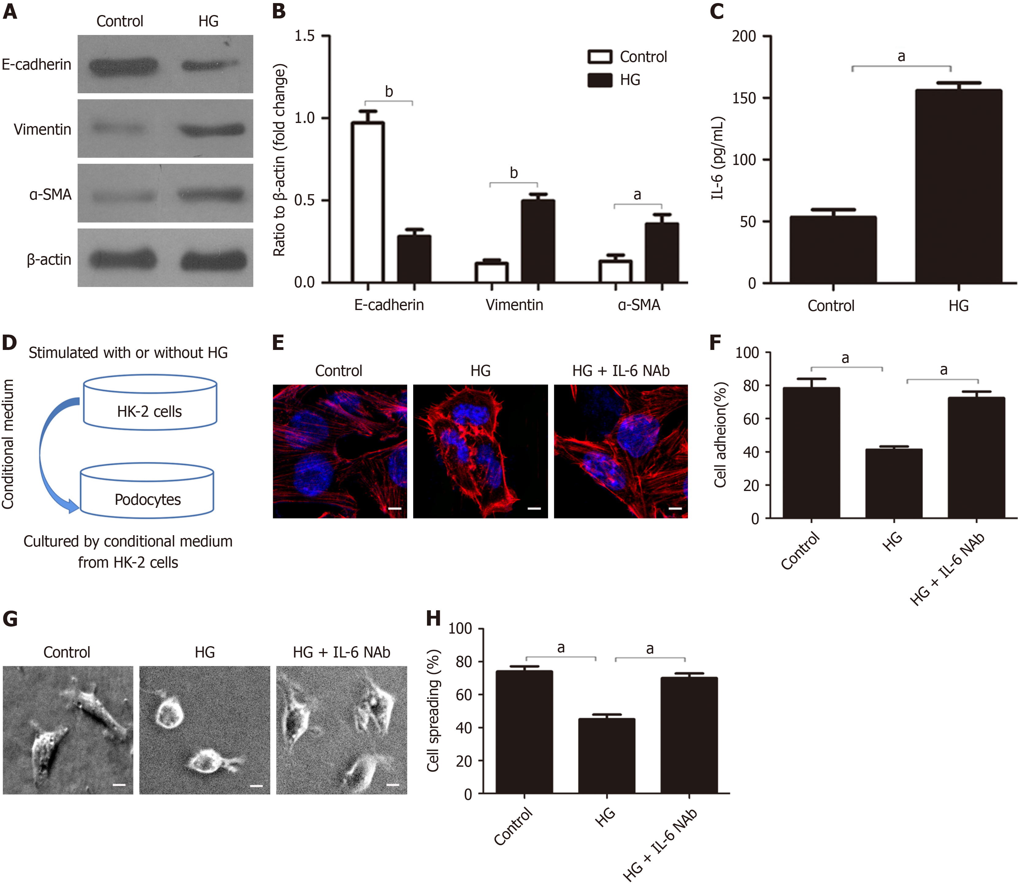
Figure 4 IL-6 secretion by HK-2 cells contributes to the HG-induced injury of podocytes.
A and B: Western blot analysis of E-cadherin, vimentin, and α-SMA in HK-2 cells after different treatments and quantification of these results (n = 3); C: ELISA measurements of IL-6 after different treatments (n = 3); D: Method used to examine the effect of HK-2 conditioned medium on podocytes; E: Representative confocal microscopy images of podocytes after culture in different conditioned media (magnification: × 1000; scale bar: 50 μm; blue: Nuclei: Red: Cytoskeleton); F: Quantitative analysis of podocyte adhesion after culture in different conditioned media (n = 3); G and H: Representative electron microscopy images of podocyte spreading after culture in different conditioned media (magnification: × 400; scale bar: 20 μm) and quantification of these results (n = 3). aP < 0.05; bP < 0.001. HG: High-dose glucose; NAb: Neutralizing Ab.
- Citation: Zha DQ, Gao P, Wu XY. Nicotinamide mononucleotide protects against diabetic nephropathy via IL-6/Rab5-mediated crosstalk between proximal tubular epithelial cells and podocytes. World J Diabetes 2025; 16(10): 109782
- URL: https://www.wjgnet.com/1948-9358/full/v16/i10/109782.htm
- DOI: https://dx.doi.org/10.4239/wjd.v16.i10.109782