
















©The Author(s) 2025.
World J Diabetes. Oct 15, 2025; 16(10): 109526
Published online Oct 15, 2025. doi: 10.4239/wjd.v16.i10.109526
Published online Oct 15, 2025. doi: 10.4239/wjd.v16.i10.109526
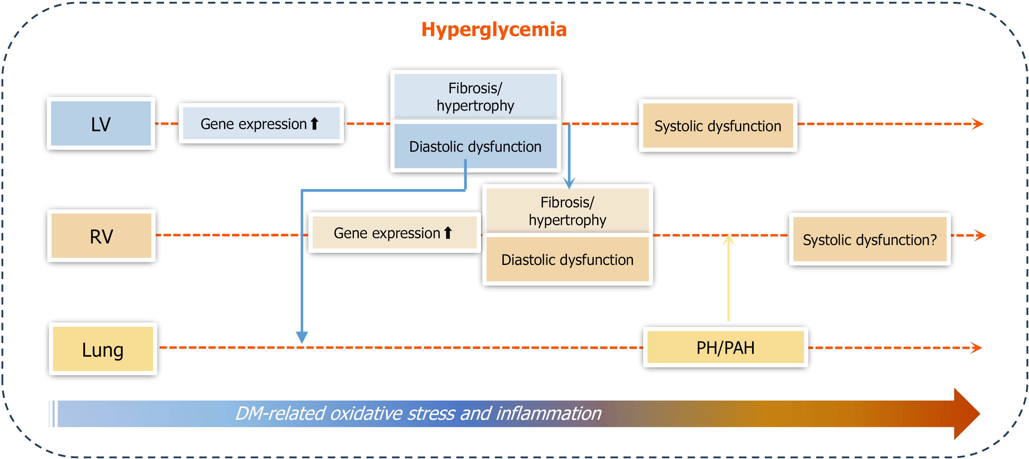
Figure 9 Outline of the main findings of the study.
Under hyperglycemic conditions that promote progressive cardiac oxidative stress and inflammation, right ventricular (RV) systolic function remained comparable to controls, whereas left ventricular (LV) systolic function declined in type 1 diabetes (T1D) mice by 30 weeks of age. RV diastolic dysfunction became evident by 18 weeks and worsened by 30 weeks, while LV diastolic dysfunction showed an increasing trend at 12 weeks, reached significance by 18 weeks, and further progressed by 30 weeks. Additionally, RV diastolic dysfunction was accompanied by RV cardiac fibrosis and hypertrophy, which occurred later than the corresponding changes in the LV. Pulmonary arterial hypertension (PAH) developed in T1D mice, evidenced by increased pulmonary acceleration time to pulmonary ejection time ratio and RV peak systolic pressure at 30 weeks. In conclusion, T1D can cause diabetic cardiomyopathy, including both RV and LV dysfunctions, which are mediated by pathological remodeling (hypertrophy and fibrosis) of each ventricle; however, diastolic dysfunction occurs earlier than systolic dysfunction. Mild PAH is present at later stages of T1D, most likely derived from LV dysfunction. The development of PAH may contribute to RV systolic impairment and remodeling. DM: Diabetes mellitus; LV: Left ventricle; RV: Right ventricle; PH: Pulmonary hypertension; PAH: Pulmonary arterial hypertension.
- Citation: Yu JJ, Han JG, Tan Y, Xu JX, LeBlanc A, Keller BB, Huang J, Cai L. Right ventricular dysfunctions in type 1 diabetic mice: A longitudinal study. World J Diabetes 2025; 16(10): 109526
- URL: https://www.wjgnet.com/1948-9358/full/v16/i10/109526.htm
- DOI: https://dx.doi.org/10.4239/wjd.v16.i10.109526