
















©The Author(s) 2025.
World J Diabetes. Oct 15, 2025; 16(10): 108714
Published online Oct 15, 2025. doi: 10.4239/wjd.v16.i10.108714
Published online Oct 15, 2025. doi: 10.4239/wjd.v16.i10.108714
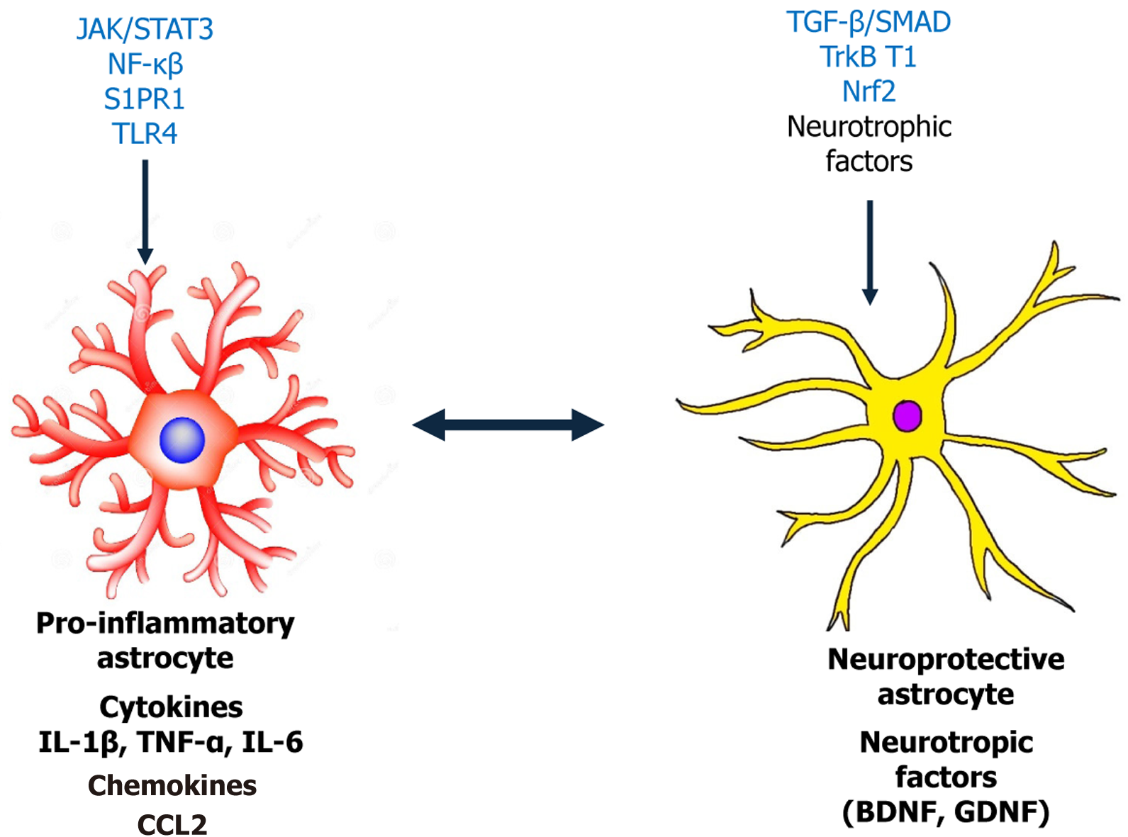
Figure 1 Functional dichotomy of glial phenotypes and signaling cascades in diabetic neuropathy.
The left panel shows pro-inflammatory astrocytes, which become reactive in response to diabetic stressors. Activation is mediated by pathways such as Janus kinase/signal transducer and activator of transcription 3 (JAK/STAT3), nuclear factor kappa B (NF-κB), Toll-like receptor 4 (TLR4), and sphingosine-1-phosphate receptor 1 (S1PR1), leading to the release of cytokines [e.g., interleukin 1 beta (IL-1β), tumor necrosis factor alpha (TNF-α), interleukin 6 (IL-6)], chemokines [e.g., C-C motif chemokine ligand 2 (CCL2)], and neurotoxic factors that promote central sensitization and neuropathic pain. The right panel illustrates neuroprotective astrocytes, driven by signaling through nuclear factor erythroid 2-related factor 2 (Nrf2), transforming growth factor beta (TGF-β)/SMAD, and tyrosine receptor kinase B (TrkB.T1), which enhance the release of neurotrophic factors [e.g., brain-derived neurotrophic factor (BDNF), glial cell line-derived neurotrophic factor (GDNF)] and antioxidants. These cells contribute to synaptic maintenance, neuronal survival, and the resolution of inflammation.
- Citation: Haris K, Long I. Role of astrocytes in diabetic neuropathy: Review of their involvement in disease mechanisms. World J Diabetes 2025; 16(10): 108714
- URL: https://www.wjgnet.com/1948-9358/full/v16/i10/108714.htm
- DOI: https://dx.doi.org/10.4239/wjd.v16.i10.108714