
















©The Author(s) 2025.
World J Diabetes. Oct 15, 2025; 16(10): 108346
Published online Oct 15, 2025. doi: 10.4239/wjd.v16.i10.108346
Published online Oct 15, 2025. doi: 10.4239/wjd.v16.i10.108346
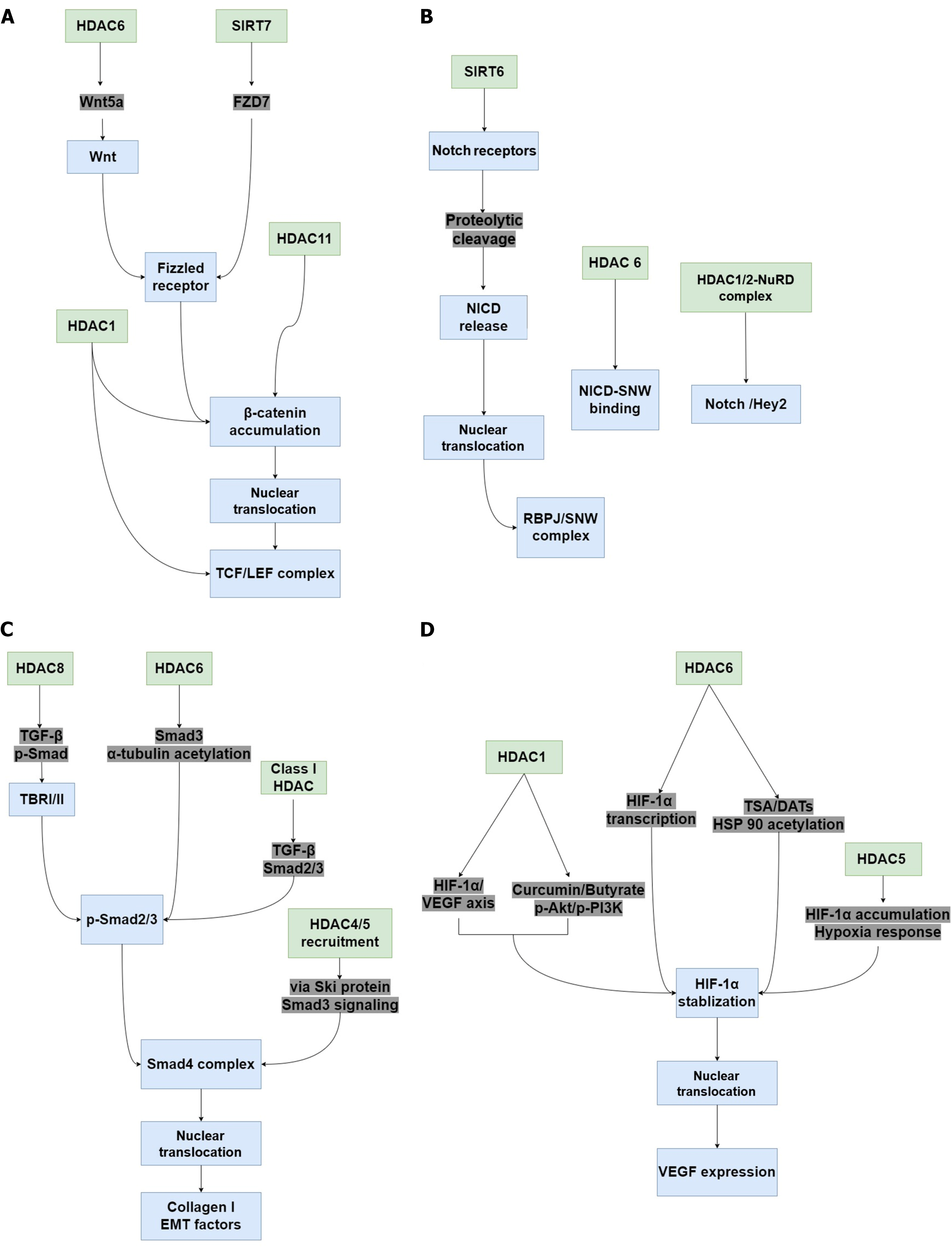
Figure 3 The potential signaling pathways associated with histone deacetylases in diabetic wound healing.
When histone deacetylase (HDAC) activity is altered in tissue, many pathways with critical roles in wound healing, such as Wnt/β-catenin, Notch, transforming growth factor (TGF)-β/suppressor of mother against decapentaplegic (Smad), and hypoxia-inducible factor-1α (HIF-1α)/vascular endothelial growth factor (VEGF) signaling are regulated. A: Inhibiting HDAC1 upregulates TCF and LEF expression, promotes the accumulation and nuclear translocation of β-catenin, and activates the Wnt/β-catenin pathway. HDAC6 increases Wnt5a expression and activates the Wnt signaling pathway. Sirtuin (SIRT) 7 promotes FZD7 expression, contributing to β-catenin stability and activation, thereby activating the Wnt/β-catenin pathway. HDAC11 suppression also promotes β-catenin stability and activates Wnt/β-catenin signaling in vascular smooth muscle cells; B: Silencing SIRT6 Leads to dysregulation of the RBPJ/SNW complex and inhibits Notch signaling. HDAC6 inhibition represses the binding of Notch intracellular cytoplasmic domain and SNW domain containing 1, thereby inhibiting Notch transcriptional responses. Recruitment of HDAC1/2-nucleosome remodeling and deacetylase also inhibits the Notch/Hey2 signaling axis; C: Inhibition of HDAC6 and HDAC8 inactivates Smad3, resulting in α-tubulin acetylation and impairing TGF-β-induced epithelial-mesenchymal transition (EMT). Class I HDAC inhibition inactivates the TGF-β-Smad2/3 signaling axis and decreases type 1 collagen expression induced by TGF-β1. The oncoprotein Sloan-kettering institute recruits HDAC4 and HDAC5, thereby inhibiting TGF-β/Smad3 signaling, leading to the decrease of EMT factors; D: Curcumin and sodium butyrate-induced HDAC1 inhibition suppresses the phospho-protein kinase B/phospho-phosphatidylinositol 3-kinase/HIF-1α/VEGF axis, whereas HDAC1 inhibition by 1,3-diphenylurea induces VEGF expression via HIF-1α, thereby increasing the migration and wound healing effects. HDAC5 overexpression enhances HIF-1α stabilization and nuclear translocation in response to hypoxia. Selective HDAC6 inhibition reduces HIF-1-mediated transcription by deacetylating heat-shock protein 90. HDAC: Histone deacetylase; SIRT: Sirtuin; NICD: Notch intracellular cytoplasmic domain; 2-NuRD: 2-nucleosome remodeling and deacetylase; TGF: Transforming growth factor; p-Smad: Phospho-suppressor of mother against decapentaplegic; TBR: Transforming growth factor-β receptor; Ski: Sloan-kettering institute; EMT: Epithelial-mesenchymal transition; VEGF: Vascular endothelial growth factor; HIF-1α: Hypoxia-inducible factor-1α; p-Akt: Phospho-protein kinase B; p-PI3K: Phospho-phosphatidylinositol 3-kinase; TSA: Trichostatin A; DATs: Diallyl trisulfide; HSP90: Heat-shock protein 90.
- Citation: Zhang F, Ma HG, Zhang B, Jiang LL, Nie KY, Deng CL, Liu Y. Potential roles of histone deacetylases in diabetic wound healing. World J Diabetes 2025; 16(10): 108346
- URL: https://www.wjgnet.com/1948-9358/full/v16/i10/108346.htm
- DOI: https://dx.doi.org/10.4239/wjd.v16.i10.108346