Copyright
©The Author(s) 2022.
World J Diabetes. Apr 15, 2022; 13(4): 338-357
Published online Apr 15, 2022. doi: 10.4239/wjd.v13.i4.338
Published online Apr 15, 2022. doi: 10.4239/wjd.v13.i4.338
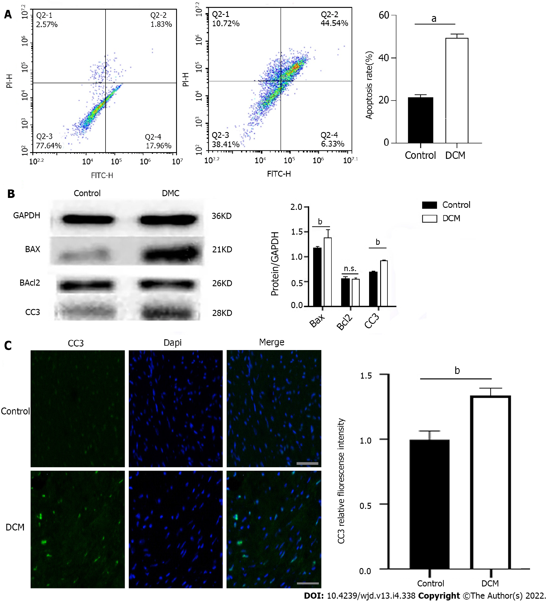
Figure 2 Apoptosis of cardiomyocytes in diabetic cardiomyopathy mice.
A: Apoptosis of diabetic cardiomyopathy (DCM) and control mice was detected using double staining of FITC-Annexin V and propidine iodide and the representative images of flow cytometry are presented. aP < 0.0001; B: Expression levels of apoptosis related proteins BAX, cleaved Caspase 3 (CC3), and Bcl2 in DCM mice and control mice. n = 5, unpaired t-test, bP < 0.01. n.s.: No statistical difference; C: Fluorescence intensity of CC3 in cardiac myocytes of DCM and control mice. Scale bars = 20 μm, bP < 0.01. DCM: Diabetic cardiomyopathy.
- Citation: Jiang SJ. Roles of transient receptor potential channel 6 in glucose-induced cardiomyocyte injury . World J Diabetes 2022; 13(4): 338-357
- URL: https://www.wjgnet.com/1948-9358/full/v13/i4/338.htm
- DOI: https://dx.doi.org/10.4239/wjd.v13.i4.338


