Copyright
©The Author(s) 2022.
World J Diabetes. Nov 15, 2022; 13(11): 972-985
Published online Nov 15, 2022. doi: 10.4239/wjd.v13.i11.972
Published online Nov 15, 2022. doi: 10.4239/wjd.v13.i11.972
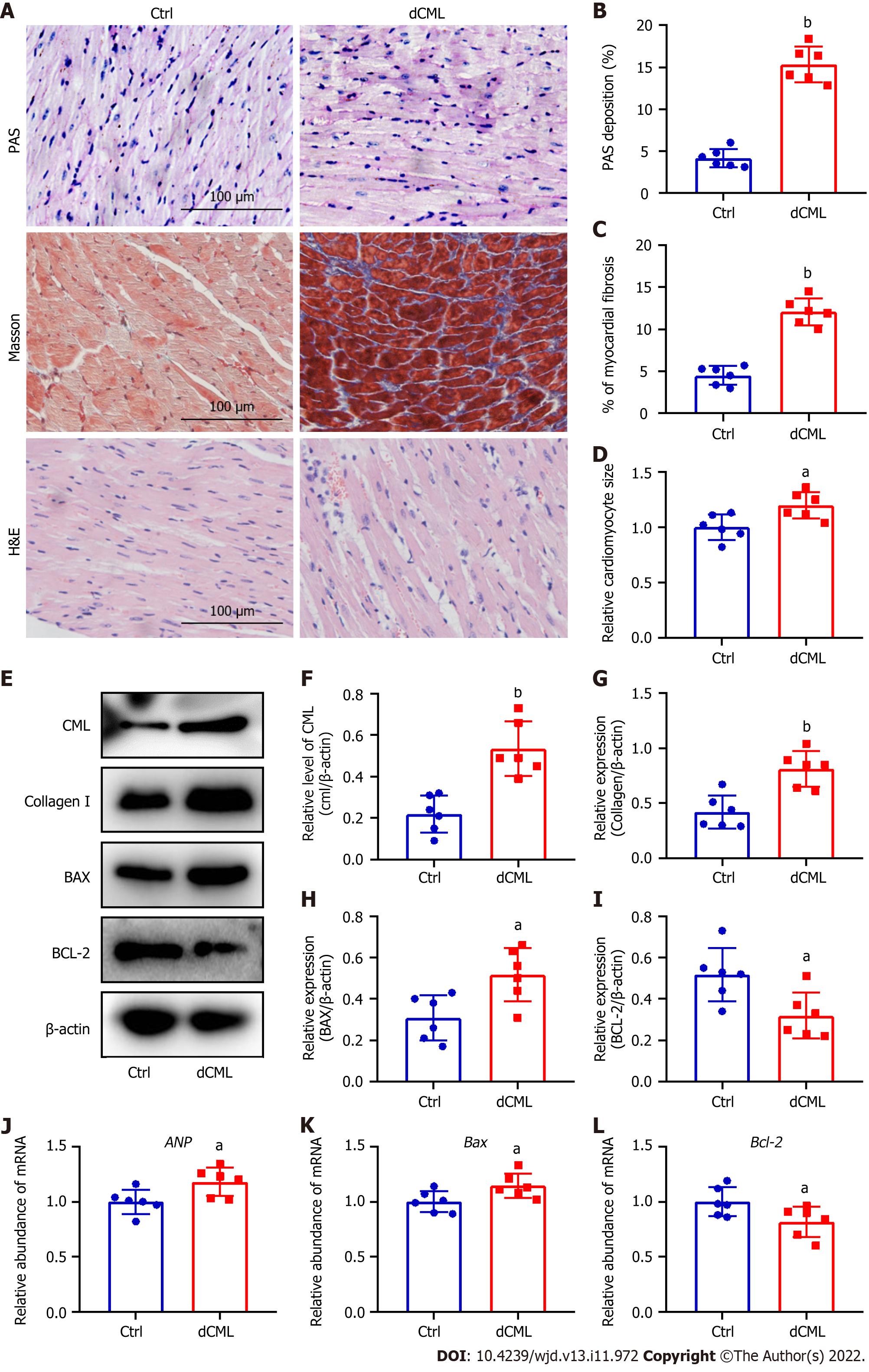
Figure 3 Dietary Nε-(carboxymethyl)lysine increases myocardial fibrosis, hypertrophy and apoptosis in mice.
A: Mouse myocardial glycogen Periodic Acid Schiff (PAS) staining, Masson’s trichrome staining, and hematoxylin and eosin staining; Scale 100 μm; B and C: Percentage of PAS-positive and fibrotic areas in the mouse myocardium; D: Relative area of myocardial cells in the myocardium; E-I: Western blotting and its relative level of Nε-(carboxymethyl)lysine (CML), collagen I, B-cell leukemia/lymphoma 2 (Bcl-2) and Bcl-2-associated X (BAX) in the mouse myocardium; J-L: Atrial natriuretic peptide (ANP), Bax, and Bcl-2 mRNA levels in the mouse myocardium. dCML: Dietary CML. n = 6. aP < 0.05, bP < 0.01, compared with the control (Ctrl) group.
- Citation: Wang ZQ, Sun Z. Dietary Nε-(carboxymethyl) lysine affects cardiac glucose metabolism and myocardial remodeling in mice. World J Diabetes 2022; 13(11): 972-985
- URL: https://www.wjgnet.com/1948-9358/full/v13/i11/972.htm
- DOI: https://dx.doi.org/10.4239/wjd.v13.i11.972


