Copyright
©The Author(s) 2024.
World J Diabetes. Feb 15, 2024; 15(2): 251-259
Published online Feb 15, 2024. doi: 10.4239/wjd.v15.i2.251
Published online Feb 15, 2024. doi: 10.4239/wjd.v15.i2.251
Figure 1 Operation of the non-mydriatic portable fundus camera when performing fundus examination.
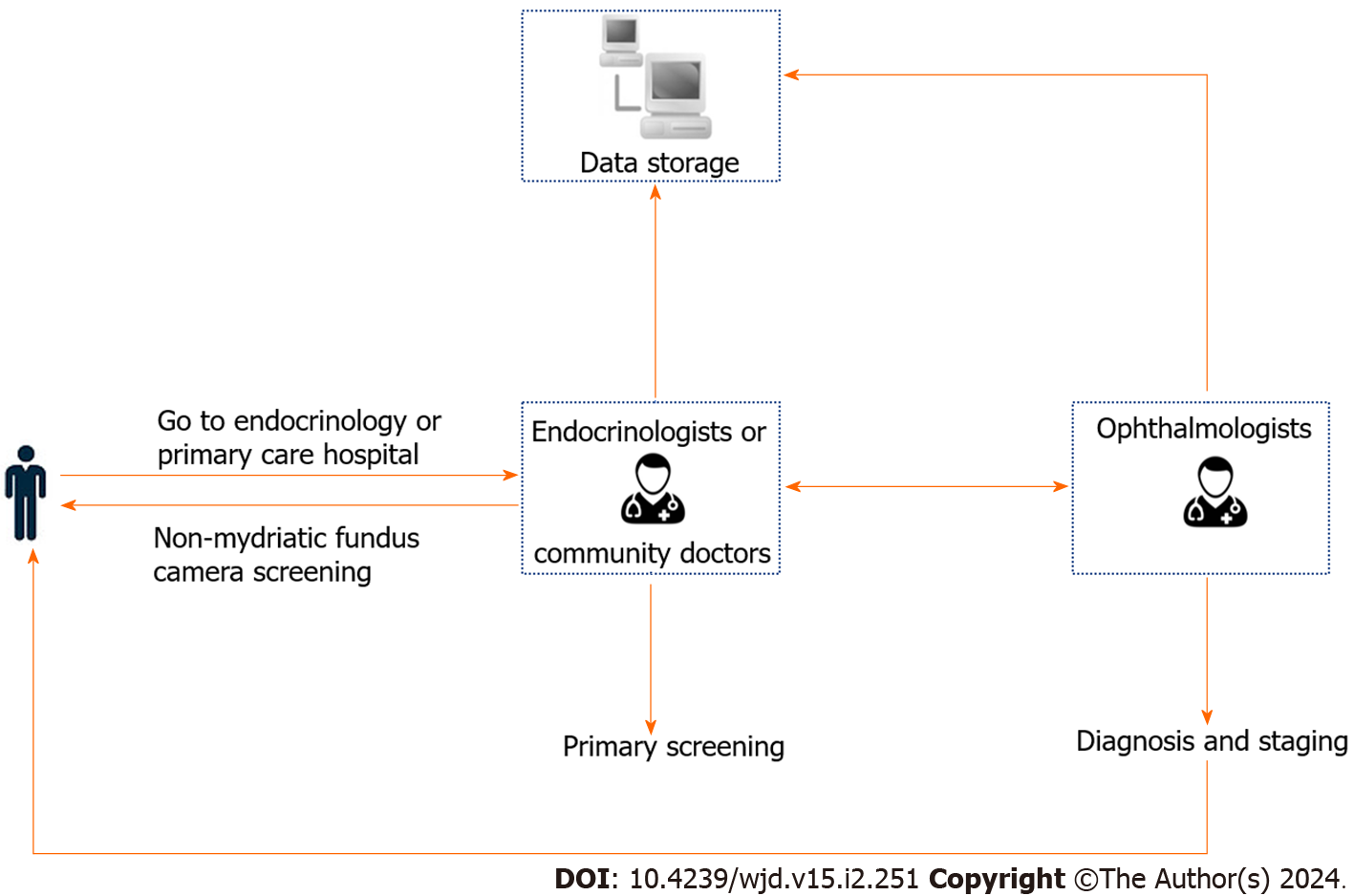
Figure 2 Application of non-mydriatic fundus photography-assisted telemedicine.
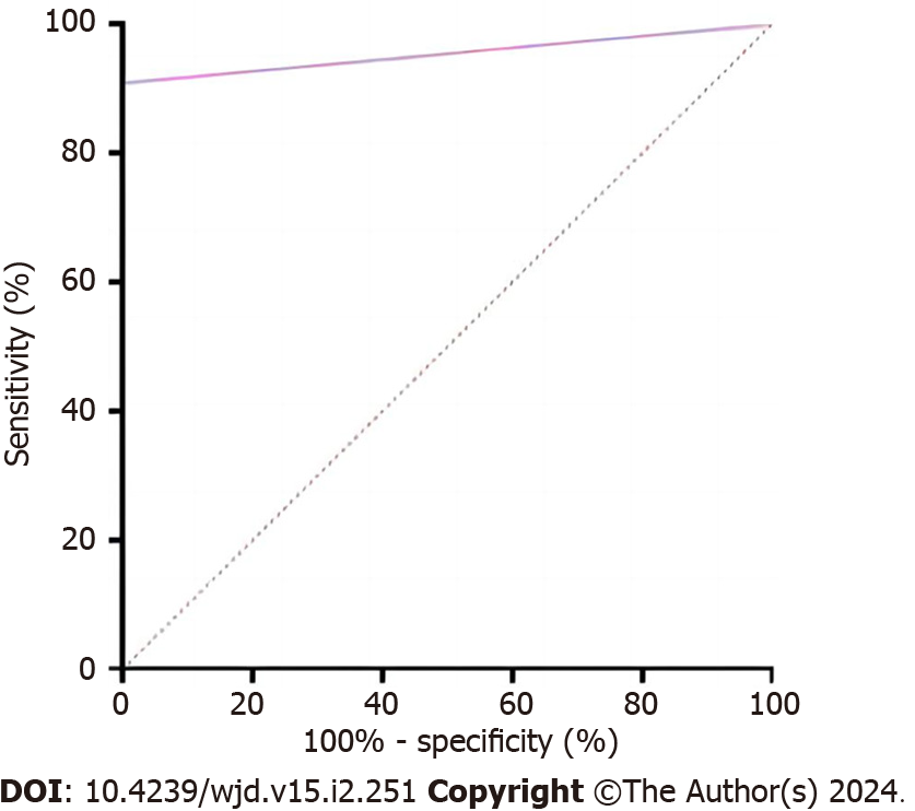
Figure 3 The receiver operating characteristic curve identified diagnostic prediction effectiveness of non-mydriatic fundus photography-assisted telemedicine (area under the curve 0.
955 with 90.9% sensitivity and 100% specificity).
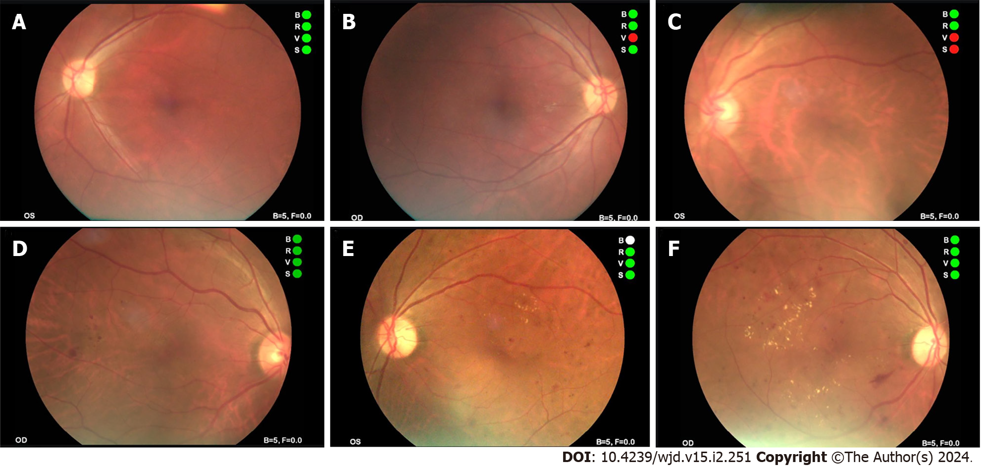
Figure 4 Typical fundus photos in different stages of non-mydriatic fundus photography-assisted telemedicine.
A and B: Photographs of normal fundus; C and D: Fundus photograph shows microhemangioma and retinal hemorrhage; E and F: Fundus photograph shows retinal hemorrhage and retinal hard exudates.
- Citation: Zhou W, Yuan XJ, Li J, Wang W, Zhang HQ, Hu YY, Ye SD. Application of non-mydriatic fundus photography-assisted telemedicine in diabetic retinopathy screening. World J Diabetes 2024; 15(2): 251-259
- URL: https://www.wjgnet.com/1948-9358/full/v15/i2/251.htm
- DOI: https://dx.doi.org/10.4239/wjd.v15.i2.251


