
















©The Author(s) 2017.
World J Gastrointest Endosc. Oct 16, 2017; 9(10): 529-534
Published online Oct 16, 2017. doi: 10.4253/wjge.v9.i10.529
Published online Oct 16, 2017. doi: 10.4253/wjge.v9.i10.529
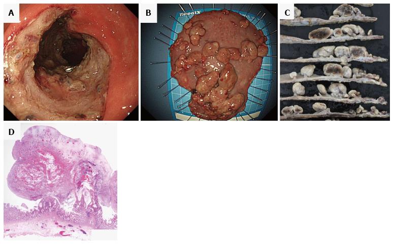
Figure 4 Endoscopic submucosal dissection.
Post-dissection ulcers after endoscopic submucosal dissection at the sigmoid colon and rectum (A). Fresh specimen of cap polyposis after endoscopic submucosal dissection (B) and fixed specimen (C). The fixed specimen revealed bleeding into the polyps. Microscopic findings of inflammatory polyps [HE stain, × 4 (D)].
- Citation: Murata M, Sugimoto M, Ban H, Otsuka T, Nakata T, Fukuda M, Inatomi O, Bamba S, Kushima R, Andoh A. Cap polyposis refractory to Helicobacter pylori eradication treated with endoscopic submucosal dissection. World J Gastrointest Endosc 2017; 9(10): 529-534
- URL: https://www.wjgnet.com/1948-5190/full/v9/i10/529.htm
- DOI: https://dx.doi.org/10.4253/wjge.v9.i10.529