Copyright
©The Author(s) 2015.
World J Gastrointest Endosc. May 16, 2015; 7(5): 496-509
Published online May 16, 2015. doi: 10.4253/wjge.v7.i5.496
Published online May 16, 2015. doi: 10.4253/wjge.v7.i5.496
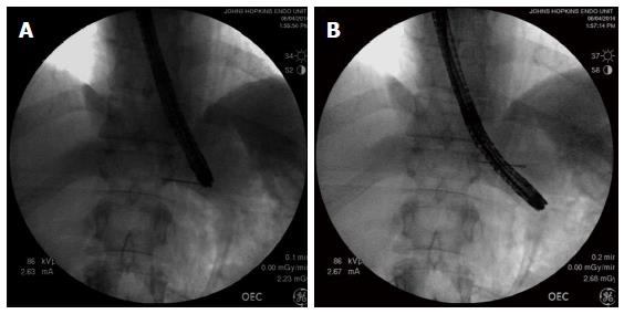
Figure 8 Using fluoroscopy to assess the adequacy of gastric myotomy during peroral endoscopic myotomy.
A: The needle is fluoroscopically lined up with the tip of the endoscope and leveled with the EGJ; B: The endoscope has a diameter of 1 cm, and therefore, the endoscope tip is measured to be 3 cm below the needle marking the EGJ. EGJ: Esophagogastric junction.
- Citation: Kumbhari V, Khashab MA. Peroral endoscopic myotomy. World J Gastrointest Endosc 2015; 7(5): 496-509
- URL: https://www.wjgnet.com/1948-5190/full/v7/i5/496.htm
- DOI: https://dx.doi.org/10.4253/wjge.v7.i5.496


