Copyright
©The Author(s) 2015.
World J Gastrointest Endosc. Mar 16, 2015; 7(3): 290-294
Published online Mar 16, 2015. doi: 10.4253/wjge.v7.i3.290
Published online Mar 16, 2015. doi: 10.4253/wjge.v7.i3.290
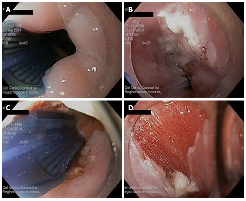
Figure 4 Radiofrequency pad is placed over the lesion under direct visualization (A); Ablation area after the first application of energy (B); Second application of the pad to include all the area of esophageal papilloma (C); Esophageal wound cleaned from debris by cleaning cup (D).
- Citation: del Genio G, del Genio F, Schettino P, Limongelli P, Tolone S, Brusciano L, Avellino M, Vitiello C, Docimo G, Pezzullo A, Docimo L. Esophageal papilloma: Flexible endoscopic ablation by radiofrequency. World J Gastrointest Endosc 2015; 7(3): 290-294
- URL: https://www.wjgnet.com/1948-5190/full/v7/i3/290.htm
- DOI: https://dx.doi.org/10.4253/wjge.v7.i3.290


