Copyright
©The Author(s) 2015.
World J Gastrointest Endosc. Mar 16, 2015; 7(3): 192-205
Published online Mar 16, 2015. doi: 10.4253/wjge.v7.i3.192
Published online Mar 16, 2015. doi: 10.4253/wjge.v7.i3.192
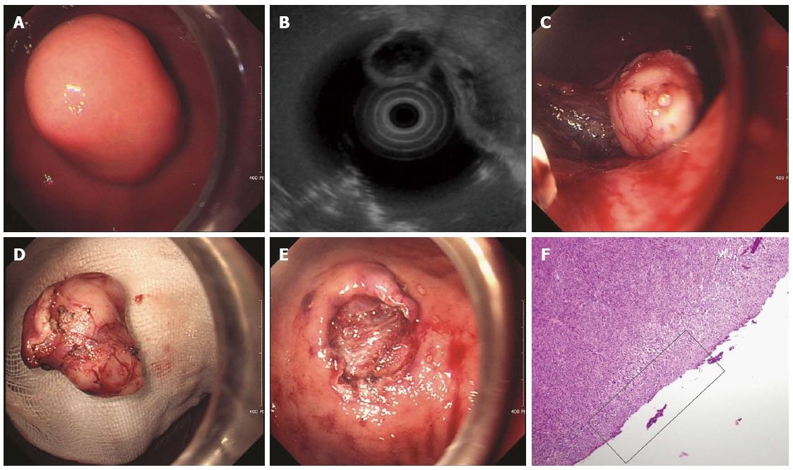
Figure 3 Endoscopic enucleation using the standard endoscopic submucosal dissection technique.
A: An approximately 2.5-cm subepithelial tumor was identified at the greater curvature side of the upper body of the stomach; B: A 2.6-cm mixed echogenic tumor with a slightly irregular border arising from the proper muscle layer was noticed; C: Endoscopic enucleation using the endoscopic submucosal dissection technique was performed; D: En bloc resection was achieved; E: There was no perforation at the operation site; F: On pathologic examination, a vertical resection margin was apparently involved with tumor cells (red boxed area); R1 resection was confirmed. (Courtesy of Kyung Oh Kim, Gil hospital, Incheon, South Korea).
- Citation: Kim HH. Endoscopic treatment for gastrointestinal stromal tumor: Advantages and hurdles. World J Gastrointest Endosc 2015; 7(3): 192-205
- URL: https://www.wjgnet.com/1948-5190/full/v7/i3/192.htm
- DOI: https://dx.doi.org/10.4253/wjge.v7.i3.192


