Copyright
©The Author(s) 2015.
World J Gastrointest Endosc. Feb 16, 2015; 7(2): 110-120
Published online Feb 16, 2015. doi: 10.4253/wjge.v7.i2.110
Published online Feb 16, 2015. doi: 10.4253/wjge.v7.i2.110
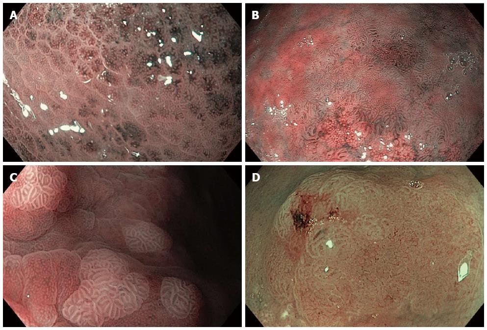
Figure 3 Narrow band imaging with magnification endoscopy images of gastric lesions.
A: Helicobacter pylori gastritis: enlargement of pits, variable vascular density (alternation of lighter and darker areas); B: Extensive areas of intestinal metaplasia (tubulovillous mucosal pattern) and remnant normal gastric body mucosa (small regular and circular pits); C: Areas of intestinal metaplasia: blue whitish slightly raised areas (the light blue crest sign) with regular, tubulovillous mucosal pattern; D: Dysplasia: area with architectural loss of mucosal pattern and irregular vascular pattern.
- Citation: Boeriu A, Boeriu C, Drasovean S, Pascarenco O, Mocan S, Stoian M, Dobru D. Narrow-band imaging with magnifying endoscopy for the evaluation of gastrointestinal lesions. World J Gastrointest Endosc 2015; 7(2): 110-120
- URL: https://www.wjgnet.com/1948-5190/full/v7/i2/110.htm
- DOI: https://dx.doi.org/10.4253/wjge.v7.i2.110


