Copyright
©The Author(s) 2015.
World J Gastrointest Endosc. Sep 25, 2015; 7(13): 1107-1113
Published online Sep 25, 2015. doi: 10.4253/wjge.v7.i13.1107
Published online Sep 25, 2015. doi: 10.4253/wjge.v7.i13.1107
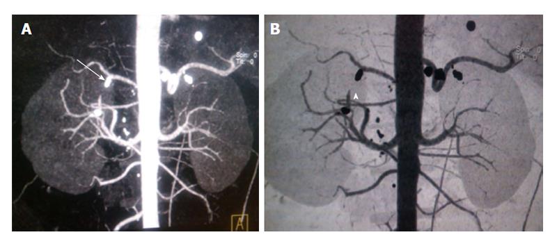
Figure 3 Follow up scan following thrombin injection procedure (case 1).
A and B: Coronal MIP (3-D reformatted) CT images. There is non-visualization of the pseudoaneurysm sac (arrowhead, B) with metallic clip (arrow, A) seen at the gastroduodenal artery stump due to previous laparoscopic surgical clipping. CT: Computed tomography; MIP: Maximum intensity projection.
- Citation: Gamanagatti S, Thingujam U, Garg P, Nongthombam S, Dash NR. Endoscopic ultrasound guided thrombin injection of angiographically occult pancreatitis associated visceral artery pseudoaneurysms: Case series. World J Gastrointest Endosc 2015; 7(13): 1107-1113
- URL: https://www.wjgnet.com/1948-5190/full/v7/i13/1107.htm
- DOI: https://dx.doi.org/10.4253/wjge.v7.i13.1107


