
















©2014 Baishideng Publishing Group Inc.
World J Gastrointest Endosc. Aug 16, 2014; 6(8): 385-389
Published online Aug 16, 2014. doi: 10.4253/wjge.v6.i8.385
Published online Aug 16, 2014. doi: 10.4253/wjge.v6.i8.385
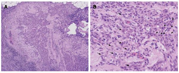
Figure 5 The surgically dissected lymph node from the neck area.
A: In the lymph node, which was 5 mm in diameter, a metastasis that was 2 mm in diameter was found (HE staining, original magnification × 40); B: Histological evaluation showed that lymphocytes had infiltrated around the carcinoma cells (HE staining, original magnification × 200). Many denatured carcinoma cells, which had karyorrhexis, an indistinct cell membrane, or karyotheca in the lymph node, were observed (arrow). Many viable carcinoma cells were observed (arrowhead).
- Citation: Uesato M, Kono T, Shiratori T, Akutsu Y, Hoshino I, Murakami K, Horibe D, Maruyama T, Semba Y, Urahama R, Ogura Y, Oide T, Tanizawa T, Matsubara H. Lymphoepithelioma-like esophageal carcinoma with macroscopic reduction. World J Gastrointest Endosc 2014; 6(8): 385-389
- URL: https://www.wjgnet.com/1948-5190/full/v6/i8/385.htm
- DOI: https://dx.doi.org/10.4253/wjge.v6.i8.385