Copyright
©2014 Baishideng Publishing Group Co.
World J Gastrointest Endosc. Jan 16, 2014; 6(1): 20-26
Published online Jan 16, 2014. doi: 10.4253/wjge.v6.i1.20
Published online Jan 16, 2014. doi: 10.4253/wjge.v6.i1.20
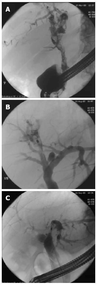
Figure 3 Endoscopic retrograde cholangiography fluoroscopic images and video illustrations of different cases.
A: Patient with multiple papillary bile duct polyps; histology revealed adenoma with high-grade intraepithelial neoplasia, liver transplantation was successfully performed; B: Patient with intrahepatic stones (S7/8) treated by laser lithotripsy; C: Patient with stenosis of the left hepatic duct; histology revealed cholangiocellular carcinoma. Peroral cholangioscopy revealed that the right hepatic duct was without signs of infiltration, and left hemihepatectomy was successfully performed.
- Citation: Prinz C, Weber A, Goecke S, Neu B, Meining A, Frimberger E. A new peroral mother-baby endoscope system for biliary tract disorders. World J Gastrointest Endosc 2014; 6(1): 20-26
- URL: https://www.wjgnet.com/1948-5190/full/v6/i1/20.htm
- DOI: https://dx.doi.org/10.4253/wjge.v6.i1.20


