Copyright
©2013 Baishideng Publishing Group Co.
World J Gastrointest Endosc. Sep 16, 2013; 5(9): 446-449
Published online Sep 16, 2013. doi: 10.4253/wjge.v5.i9.446
Published online Sep 16, 2013. doi: 10.4253/wjge.v5.i9.446
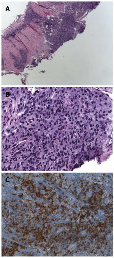
Figure 2 Photographs from the hematoxylin and eosin stain, and immunohistochemistry staining, of the biopsy samples.
A: Photomicrograph shows atypical infiltrate under the mucosa of the esophagus × 40 original magnification hematoxylin and eosin (HE) stain; B: The tumor cells are medium sized lymphocytes and have a round or slightly constricted nuclei, × 400 original magnification, HE stain; C: Immunohistochemistry: CD20, CD3, CD43, Kappa and Lambda.
- Citation: Malik AO, Baig Z, Ahmed A, Qureshi N, Malik FN. Extremely rare case of primary esophageal mucous associated lymphoid tissue lymphoma. World J Gastrointest Endosc 2013; 5(9): 446-449
- URL: https://www.wjgnet.com/1948-5190/full/v5/i9/446.htm
- DOI: https://dx.doi.org/10.4253/wjge.v5.i9.446


