
















©2013 Baishideng Publishing Group Co.
World J Gastrointest Endosc. Mar 16, 2013; 5(3): 128-131
Published online Mar 16, 2013. doi: 10.4253/wjge.v5.i3.128
Published online Mar 16, 2013. doi: 10.4253/wjge.v5.i3.128
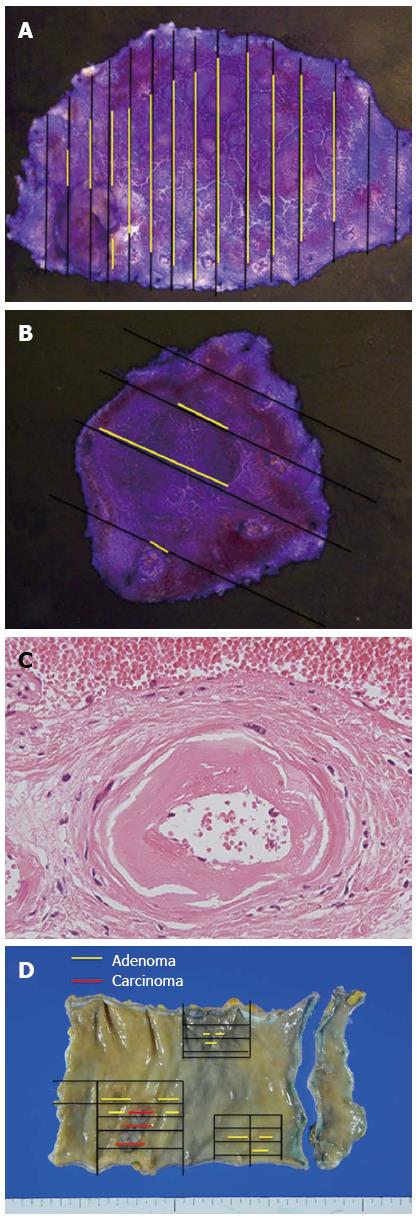
Figure 4 Resected specimens.
A: A complete one-piece resection of 52 mm × 30 mm in size with tumor-free margins was achieved; B: Complete one-piece resection with tumor-free margins 18 mm × 18 mm; C: Microscopy of the resected specimens revealed increased vessel wall thickness in the submucosal layer and the serous coat of the large intestine around the tumors, indicating chronic radiation proctitis; D: A well-differentiated adenocarcinoma, 35 mm × 18 mm, invading beyond the muscularis propriae (pT3). None of the 10 lymph nodes retrieved were involved (pN0). Moreover, adenomatous change, which endoscopic observation failed to detect, was found.
- Citation: Asayama N, Ikehara H, Yano H, Saito Y. Endoscopic submucosal dissection of multiple flat adenomas in the radiated rectum. World J Gastrointest Endosc 2013; 5(3): 128-131
- URL: https://www.wjgnet.com/1948-5190/full/v5/i3/128.htm
- DOI: https://dx.doi.org/10.4253/wjge.v5.i3.128