Copyright
©2013 Baishideng Publishing Group Co.
World J Gastrointest Endosc. Nov 16, 2013; 5(11): 581-583
Published online Nov 16, 2013. doi: 10.4253/wjge.v5.i11.581
Published online Nov 16, 2013. doi: 10.4253/wjge.v5.i11.581
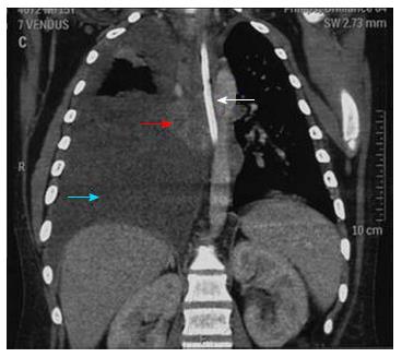
Figure 2 Computerised tomography of the thorax showing thickening of the mid-esophagus (red arrow) along with Ryle’s tube in situ (white arrow).
Right sided pleural effusion seen (blue arrow).
- Citation: Jain SS, Somani PO, Mahey RC, Shah DK, Contractor QQ, Rathi PM. Esophageal tuberculosis presenting with hematemesis. World J Gastrointest Endosc 2013; 5(11): 581-583
- URL: https://www.wjgnet.com/1948-5190/full/v5/i11/581.htm
- DOI: https://dx.doi.org/10.4253/wjge.v5.i11.581


