Copyright
©2012 Baishideng.
World J Gastrointest Endosc. Aug 16, 2012; 4(8): 356-361
Published online Aug 16, 2012. doi: 10.4253/wjge.v4.i8.356
Published online Aug 16, 2012. doi: 10.4253/wjge.v4.i8.356
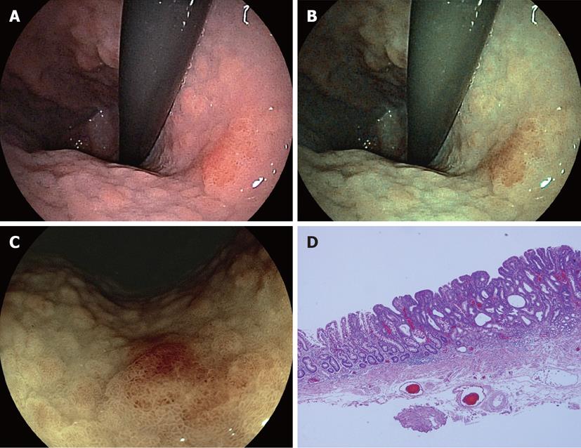
Figure 1 Image findings and specimen, A: Conventional image with small caliber endoscope (EG530-NW) reveals a slightly reddish mucosal change in the lesser curvature of the upper body; B: Flexible spectral imaging color enhancement (FICE) image with small caliber endoscope enhances a reddish cancerous lesion and can determine with precision a clear line of demarcation between cancer and the yellowish surrounding mucosa; C: FICE image with low magnification (EG590-ZW) also detects much clearer demarcation line; D: Specimen after endoscopic submucosal dissection shows a high density of glandular structure and an apparently irregular microvessel in intervening parts between crypts, which may cause a reddish mucosal change in depressed area.
- Citation: Osawa H, Yamamoto H, Miura Y, Yoshizawa M, Sunada K, Satoh K, Sugano K. Diagnosis of extent of early gastric cancer using flexible spectral imaging color enhancement. World J Gastrointest Endosc 2012; 4(8): 356-361
- URL: https://www.wjgnet.com/1948-5190/full/v4/i8/356.htm
- DOI: https://dx.doi.org/10.4253/wjge.v4.i8.356


