Copyright
©2012 Baishideng Publishing Group Co.
World J Gastrointest Endosc. Dec 16, 2012; 4(12): 565-570
Published online Dec 16, 2012. doi: 10.4253/wjge.v4.i12.565
Published online Dec 16, 2012. doi: 10.4253/wjge.v4.i12.565
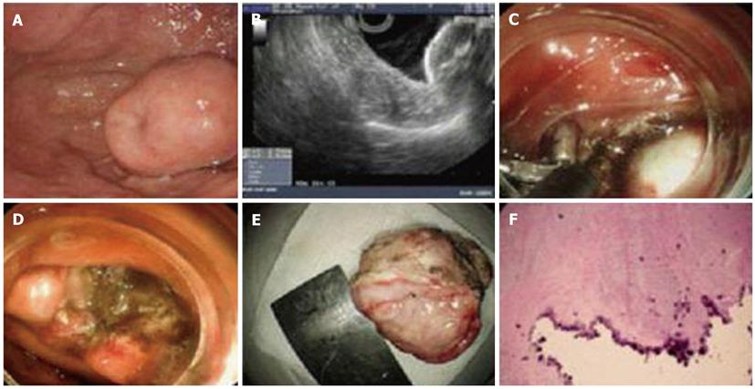
Figure 3 Gastrointestinal stromal tumors in the fundus were dissected by endoscopy.
A: Submucosal tumor located in the fundus; B: The lesion was shown in the muscularis propria by endoscopic ultrasonography (EUS); C: White lesion was clearly identified, two clips were used to arrest bleeding from small submucosal vessel; D: The wound surface after dissection of the lesion; E: The dissected tumor (3.0 cm × 2.0 cm) with intact envelope; F: Pathological image of the resected gastrointestinal stromal tumors and calcification in the center of the lesion in accordance with EUS image.
- Citation: Huang ZG, Zhang XS, Huang SL, Yuan XG. Endoscopy dissection of small stromal tumors emerged from the muscularis propria in the upper gastrointestinal tract: Preliminary study. World J Gastrointest Endosc 2012; 4(12): 565-570
- URL: https://www.wjgnet.com/1948-5190/full/v4/i12/565.htm
- DOI: https://dx.doi.org/10.4253/wjge.v4.i12.565


