Copyright
©2011 Baishideng Publishing Group Co.
World J Gastrointest Endosc. Jun 16, 2011; 3(6): 110-117
Published online Jun 16, 2011. doi: 10.4253/wjge.v3.i6.110
Published online Jun 16, 2011. doi: 10.4253/wjge.v3.i6.110
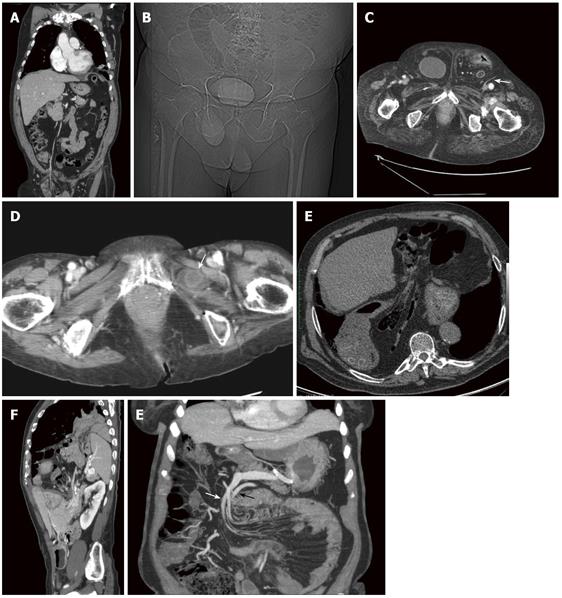
Figure 2 Computed tomography.
A: Coronal reconstruction of a right indirect inguinal hernia. Bowel loops are visible in the hernial sac and the vascular axis passing trough the inguinal canal; B: Pilot scan. Bladder underlined by the contrast media in a right direct inguinal hernia; C: Axial scan. Bilateral direct inguinal hernia. On the right contains the bladder, on the left intestinal loops. Note the epigastric vessels lateral to the hernia (arrow); D: Obturator hernias. A thickened bowel loop is located between the external obturator and pectineal muscles (arrow); E: Very large hiatal hernia containing also mesenteric fat and bowel loops; F: Sagittal reconstruction of bockdaleck hernia. The spleen, part of the left kidney and small bowel pass in the thorax through a posterior diaphragmatic defect; G: Thick slab MIP coronal reconstruction of left paraduodenal hernia. Both, the inferior mesenteric vein (white arrow) and the ascending left colic artery (black arrow) can be seen above the herniated loop along the anterior aspect.
- Citation: Lassandro F, Iasiello F, Pizza NL, Valente T, Stefano MLMDS, Grassi R, Muto R. Abdominal hernias: Radiological features. World J Gastrointest Endosc 2011; 3(6): 110-117
- URL: https://www.wjgnet.com/1948-5190/full/v3/i6/110.htm
- DOI: https://dx.doi.org/10.4253/wjge.v3.i6.110


